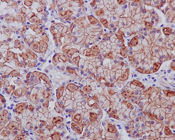
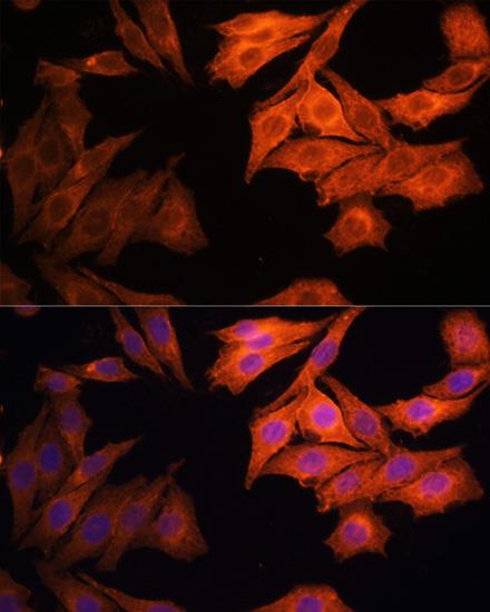
HER1 / EGFR
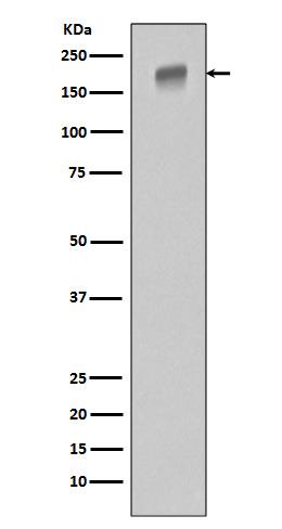
Western blot analysis of extracts of OVCAR3 cells, using EGFR Rabbit pAb at 1:1000 dilution. Secondary antibody: HRP Goat Anti-Rabbit IgG (H+L) at 1:10000 dilution. Lysates/proteins: 25ug per lane. Blocking buffer: 3% nonfat dry milk in TBST. Detection: ECL West Pico Plus. Exposure time: 10s.
| Catalog Number: | MAB-94446 |
| Conjugate: | unconjugated |
| Size: | 100 ug |
| Concentration: | 1mg/ml |
| Host: | Ms |
| Isotype: | IgG2b |
| Clone: | IS-23 |
| Immunogen: | A synthesized peptide derived from human EGFR (ErbB 1) Antibody EGFR is a receptor tyrosine kinase. Receptor for epidermal growth factor (EGF) and related growth factors including TGFalpha, amphiregulin, betacellulin, heparin-binding EGF-like growth factor, GP30 and vaccinia virus growth factor. Is involved in the control of cell growth and differentiation. |
| Reactivity: | Hu, Ms, Rt |
| Applications: | Western Blot: 1:500-1:2000 Immunohistochemistry: 1:50-1:100 Immunocytochemistry: 1:50- 1:200 Immunofluorescence: 1:50-1:200 Immunoprecipitation: 1:50-1:200 |
| Molecular Weight: | 175kDa |
| Purification: | Affinity-chromatography |
| Form: | Liquid |
| Synonyms: | Epidermal Growth Factor Receptor ;ErbB-1; ErbB1; ER1; ERB-B1; MENA; HER1; Erythroblastic Leukemia Viral(v-Erb-B)oncogene HomologAvian; Proto-oncogene c-ErbB-1; Receptor tyrosine-protein kinase erbB-1 |
The epidermal growth factor receptor (EGFR; ErbB-1; HER1 in humans) is the cell-surface receptor for members of the epidermal growth factor family (EGF-family) of extracellular protein ligands. Known ligands include EGF, TGFA/TGF-alpha, amphiregulin, epigen/EPGN, BTC/betacellulin, epiregulin/EREG and HBEGF/heparin-binding EGF. Ligand binding triggers receptor homo- and/or heterodimerization and autophosphorylation on key cytoplasmic residues. The phosphorylated receptor recruits adapter proteins like GRB2 which in turn activates complex downstream signaling cascadesβ-Actin is one of six different actin isoforms that have been identified. The actin molecules found in cells of various species and tissues tend to be very similar in their immunological and physical properties. Therefore, Antibodies against β-Actin are useful as loading controls for Western Blotting. However it should be noted that levels of β-Actin may not be stable in certain cells. For example, expression of β-Actin in adipose tissue is very low and therefore β-Actin should not be used as loading control for these tissues.
Applications
Western Blot: 1:500-1:2000 Immunohistochemistry: 1:50-1:100 Immunocytochemistry: 1:50- 1:200 Immunofluorescence: 1:50-1:200 Immunoprecipitation: 1:50-1:200
Immunogen
A synthesized peptide derived from human EGFR (ErbB 1) Antibody EGFR is a receptor tyrosine kinase. Receptor for epidermal growth factor (EGF) and related growth factors including TGFalpha, amphiregulin, betacellulin, heparin-binding EGF-like growth factor, GP30 and vaccinia virus growth factor. Is involved in the control of cell growth and differentiation.