
HIF-1alpha

Western blot analysis of extracts of
mouse kidney, using HIF1α antibody
at 1:1000 dilution.
Secondary antibody: HRP Goat Anti-
Rabbit IgG (H+L) at 1:10000
dilution.
Lysates/proteins: 25ug per lane.
Blocking buffer: 3% nonfat dry milk in
TBST.
Detection: ECL West Pico Plus. 
Exposure time: 5s.

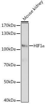
Western blot analysis of extracts of mouse kidney, using HIF1α antibody at 1:1000 dilution. Secondary antibody: HRP Goat Anti- Rabbit IgG (H+L) at 1:10000 dilution. Lysates/proteins: 25ug per lane. Blocking buffer: 3% nonfat dry milk in TBST. Detection: ECL West Pico Plus. Exposure time: 5s.

Catalog Number:AB-84304
Conjugate:Unconjugated
Size:100 ug
Concentration:1mg/ml
Host:Rb
Isotype:IgG
Clone:POLY
Immunogen:Recombinant protein of human HIF1α.
Reactivity:Hu, Ms, Rt
Applications:Western Blot: 1:500 - 1:2000 Immunohistochemistry: 1:50 - 1:200
Molecular Weight:110kDa
Purification:Aff. Pur.
Form:Liquid
Synonyms:HIF1A;HIF-1-alpha;HIF-1A;HIF-1alpha;HIF1;HIF1-ALPHA;MOP1;PASD8;bHLHe78

Applications

Western Blot: 1:500 - 1:2000 Immunohistochemistry: 1:50 - 1:200

Immunogen

Recombinant protein of human HIF1α.
Target Background

This gene encodes the alpha subunit of transcription factor hypoxia-inducible factor-1 (HIF-1), which is a heterodimer composed of an alpha and a beta subunit. HIF-1 functions as a master regulator of cellular and systemic homeostatic response to hypoxia by activating transcription of many genes, including those involved in energy metabolism, angiogenesis, apoptosis, and other genes whose protein products increase oxygen delivery or facilitate metabolic adaptation to hypoxia. HIF-1 thus plays an essential role in embryonic vascularization, tumor angiogenesis and pathophysiology of ischemic disease. Alternatively spliced transcript variants encoding different isoforms have been identified for this gene.

Synonyms:HIF1A;HIF-1-alpha;HIF-1A;HIF-1alpha;HIF1;HIF1-ALPHA;MOP1;PASD8;bHLHe78

Storage

Store at -20°C, and avoid repeat freeze-thaw cycles.

Buffer

PBS with 0.02% sodium azide, 50% glycerol, pH7.3.

Western blot analysis of extracts of mouse kidney, using HIF1α antibody at 1:1000 dilution. Secondary antibody: HRP Goat Anti- Rabbit IgG (H+L) at 1:10000 dilution. Lysates/proteins: 25ug per lane. Blocking buffer: 3% nonfat dry milk in TBST. Detection: ECL West Pico Plus. Exposure time: 5s.

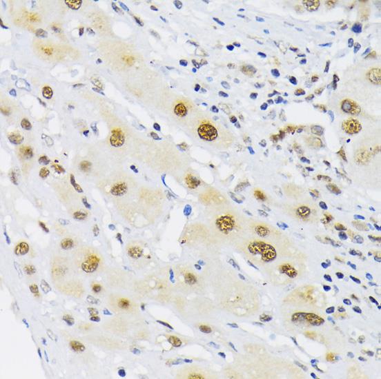
Immunohistochemistry of paraffin Embedded human liver cancer using HIF1α antibody at dilution of 1:200 (40x lens).

No documents available.