HIF-1alpha

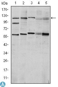
Western Blot (WB) analysis using HIF-1alpha Monoclonal Antibody against Cos7 (1), HeLa (2), Jurkat (3), RAJI (4) and NIH/3T3 (5) cell lysate.

Western Blot (WB) analysis using HIF-1alpha Monoclonal Antibody against Cos7 (1), HeLa (2), Jurkat (3), RAJI (4) and NIH/3T3 (5) cell lysate.

Catalog Number:MAB-94581
Conjugate:Unconjugated
Size:100 ug
Concentration:1mg/ml
Host:Ms
Isotype:IgG1
Clone:1A3
Immunogen:Purified recombinant fragment of human HIF-1alpha expressed in E. Coli
Reactivity:Hu, Ms, Sm
Applications:Western Blot: 1:500-1:2000 Immunohistochemistry: 1:200-1:1000 Immunofluorescence: 1:200-1:1000 ELISA: 1:10000
Purification:Aff. Pur.
Form:Liquid
Synonyms:Hypoxia-inducible factor 1-alpha HIF-1-alpha HIF1-alpha ARNT-interacting protein Basic-helix-loop-helix-PAS protein MOP1 Class E basic helix-loophelix protein 78 bHLHe78 Member of PAS protein 1 PAS domain-c

Applications

Western Blot: 1:500-1:2000 Immunohistochemistry: 1:200-1:1000 Immunofluorescence: 1:200-1:1000 ELISA: 1:10000

Immunogen

Purified recombinant fragment of human HIF-1alpha expressed in E. Coli
Target Background

Specificity: HIF-1alpha Monoclonal Antibody detects endogenous levels of HIF-1alpha protein. Tissue Specificity: Expressed in most tissues with highest levels in kidney and heart. Overexpressed in the majority of common human cancers and their metastases, due to the presence of intratumoral hypoxia and as a result of mutations in genes encoding oncoproteins and tumor suppressors. A higher level expression seen in pituitary tumors as compared to the pituitary gland.

Synonyms:Hypoxia-inducible factor 1-alpha HIF-1-alpha HIF1-alpha ARNT-interacting protein Basic-helix-loop-helix-PAS protein MOP1 Class E basic helix-loophelix protein 78 bHLHe78 Member of PAS protein 1 PAS domain-c

Storage

Store at -20°C, and avoid repeat freeze-thaw cycles.

Buffer

Ascitic fluid containing 0.03% sodium azide.

Western Blot (WB) analysis using HIF-1alpha Monoclonal Antibody against Cos7 (1), HeLa (2), Jurkat (3), RAJI (4) and NIH/3T3 (5) cell lysate.

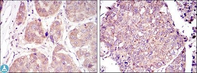
Immunohistochemistry (IHC) analysis of paraffin-embedded liver cancer tissues (left) and Lung Cancer Tissues (right) with DAB staining using HIF-1alpha Monoclonal Antibody.

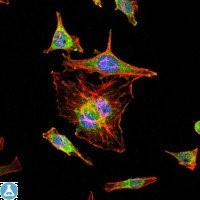
Immunofluorescence (IF) analysis of HeLa cells using HIF-1alpha Monoclonal Antibody (green). Blue: DRAQ5 fluorescent DNA dye. Red: Actin filaments have been labeled with Alexa Fluor-555 phalloidin.

No documents available.