IGF1R Rabbit Polyclonal Antibody
![Western blot analysis of lysates from wild type (WT) and IGF1R knockdown (KD) 293T cells using
[KDValidated] IGF1R Rabbit pAb at 1:3000 dilution.
Secondary antibody:HRP Goat Anti-Rabbit IgG (H+L) at 1:10000 dilution.
Lysates/proteins: 25 μg per lane.
Blocking buffer: 3% nonfat dry milk in TBST.
Detection: ECL West Pico Plus.
Exposure time: 10s.](/storage/products/wp-images/2022/01/AB-81364-1.jpg)
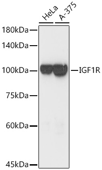
Western blot analysis of lysates from wild type (WT) and IGF1R knockdown (KD) 293T cells using [KDValidated] IGF1R Rabbit pAb at 1:3000 dilution. Secondary antibody:HRP Goat Anti-Rabbit IgG (H+L) at 1:10000 dilution. Lysates/proteins: 25 μg per lane. Blocking buffer: 3% nonfat dry milk in TBST. Detection: ECL West Pico Plus. Exposure time: 10s.
| Catalog Number: | AB-81364 |
| Conjugate: | Unconjugated |
| Size: | 100μg |
| Concentration: | 1mg/ml |
| Host: | Rabbit |
| Isotype: | IgG |
| Clone: | POLY |
| Immunogen: | Recombinant fusion protein containing a sequence corresponding to amino acids 1201-1367 of human IGF1R. |
| Reactivity: | Human,Mouse,Rat |
| Applications: | Western Blot: 1:1000 - 1:5000 |
| Molecular Weight: | 100kDa |
| Purification: | Affinity purification |
| Form: | Liquid |
| Synonyms: | IGFR; CD221; IGFIR; JTK13; 1R |
Applications
Western Blot: 1:1000 - 1:5000
Immunogen
Recombinant fusion protein containing a sequence corresponding to amino acids 1201-1367 of human IGF1R.