IL-1β Rabbit Monoclonal Antibody

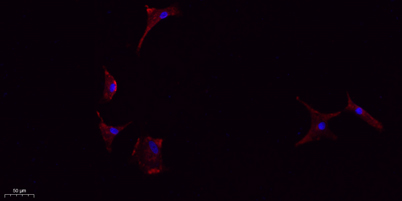
Immunofluorescence analysis of A549. 1,primary Antibody (red) was diluted at 1:200 (4°C overnight). 2, Goat Anti Rabbit IgG (H&L) - Alexa Fluor 594 Secondary antibody was diluted at 1:1000 (room temperature, 50min).3, Picture B: DAPI (blue) 10min.

Immunofluorescence analysis of A549. 1,primary Antibody (red) was diluted at 1:200 (4°C overnight). 2, Goat Anti Rabbit IgG (H&L) - Alexa Fluor 594 Secondary antibody was diluted at 1:1000 (room temperature, 50min).3, Picture B: DAPI (blue) 10min.

Catalog Number:MAB-94634
Conjugate:unconjugated
Size:100 ug
Concentration:1 mg/ml
Host:Rabbit
Isotype:IgG
Clone:MI1
Immunogen:The antiserum was produced against synthesized peptide derived from the Internal region of human IL1B. AA range:181-230
Reactivity:Human;Mouse;Rat
Applications:Western Blot: 1/500 - 1/2000. IHC-p: 1:100-300 ELISA: 1/20000. IF 1:100-300 Not yet tested in other applications.
Molecular Weight:17kD
Form:liquid

The protein encoded by this gene is a member of the interleukin 1 cytokine family. This cytokine is produced by activated macrophages as a proprotein, which is proteolytically processed to its active form by caspase 1 (CASP1/ICE). This cytokine is an important mediator of the inflammatory response, and is involved in a variety of cellular activities, including cell proliferation, differentiation, and apoptosis. The induction of cyclooxygenase-2 (PTGS2/COX2) by this cytokine in the central nervous system (CNS) is found to contribute to inflammatory pain hypersensitivity. This gene and eight other interleukin 1 family genes form a cytokine gene cluster on chromosome 2. [provided by RefSeq, Jul 2008],

Applications

Western Blot: 1/500 - 1/2000. IHC-p: 1:100-300 ELISA: 1/20000. IF 1:100-300 Not yet tested in other applications.

Immunogen

The antiserum was produced against synthesized peptide derived from the Internal region of human IL1B. AA range:181-230

Target Background

The protein encoded by this gene is a member of the interleukin 1 cytokine family. This cytokine is produced by activated macrophages as a proprotein, which is proteolytically processed to its active form by caspase 1 (CASP1/ICE). This cytokine is an important mediator of the inflammatory response, and is involved in a variety of cellular activities, including cell proliferation, differentiation, and apoptosis. The induction of cyclooxygenase-2 (PTGS2/COX2) by this cytokine in the central nervous system (CNS) is found to contribute to inflammatory pain hypersensitivity. This gene and eight other interleukin 1 family genes form a cytokine gene cluster on chromosome 2. [provided by RefSeq, Jul 2008],

Storage

-20°C/1 year

Immunofluorescence analysis of A549. 1,primary Antibody (red) was diluted at 1:200 (4°C overnight). 2, Goat Anti Rabbit IgG (H&L) - Alexa Fluor 594 Secondary antibody was diluted at 1:1000 (room temperature, 50min).3, Picture B: DAPI (blue) 10min.

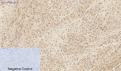
Immunohistochemical analysis of paraffin-embedded Human-uterus tissue. 1,IL-1β Polyclonal Antibody was diluted at 1:200 (4°C,overnight). 2, Sodium citrate pH 6.0 was used for antibody retrieval( >98°C,20min). 3,Secondary antibody was diluted at 1:200 (room temperature, 30min). Negative control was used by secondary antibody only.

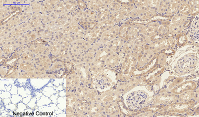
Immunohistochemical analysis of paraffin-embedded Rat-lung tissue. 1,IL-1β Polyclonal Antibody was diluted at 1:200(4°C,overnight). 2, Sodium citrate pH 6.0 was used for antibody retrieval(>98°C,20min). 3,Secondary antibody was diluted at 1:200(room temperature)

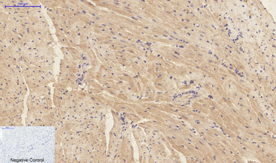
Immunohistochemical analysis of paraffin-embedded Mouse-heart tissue. 1,IL-1β Polyclonal Antibody was diluted at 1:200(4°C,overnight). 2, Sodium citrate pH 6.0 was used for antibody retrieval(>98°C,20min). 3,Secondary antibody was diluted at 1:200(room temperature 30min). Negative control was used by secondary antibody only.

No documents available.