IL-6 Rabbit Polyclonal Antibody

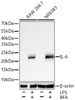
Western blot analysis of various lysates using IL-6 Rabbit PAb  at 1:5000 dilution incubated overnight at 4℃. RAW 264.7 cells and NR8383 cells were treated with LPS (1 μg/mL) and BFA (300 ng/mL) for 8 hours.
Secondary antibody: HRP-conjugated Goat anti-Rabbit IgG (H+L)  at 1:10000 dilution.
Lysates/proteins: 30 μg per lane.
Blocking buffer: 3% nonfat dry milk in TBST.
Detection: ECL West Pico Plus.
Exposure time: 45 s.

Western blot analysis of various lysates using IL-6 Rabbit PAb at 1:5000 dilution incubated overnight at 4℃. RAW 264.7 cells and NR8383 cells were treated with LPS (1 μg/mL) and BFA (300 ng/mL) for 8 hours. Secondary antibody: HRP-conjugated Goat anti-Rabbit IgG (H+L) at 1:10000 dilution. Lysates/proteins: 30 μg per lane. Blocking buffer: 3% nonfat dry milk in TBST. Detection: ECL West Pico Plus. Exposure time: 45 s.

Catalog Number:ABE4078
Conjugate:Unconjugated
Size:100 ug
Concentration:1mg/ml
Host:Rabbit
Isotype:IgG
Clone:Polyclonal
Immunogen:Recombinant protein. This information is considered to be commercially sensitive.
Reactivity:Mouse,Rat
Applications:WB 1:1000 - 1:5000 IP 0.5 μg-4 μg antibody for 900 μg-1100 μg extracts of whole cells IF/ICC 1:200 - 1:800 ELISA
Molecular Weight:24 kDa
Purification:Affinity purification
Form:liquid
Synonyms:ILg6; Ifnb2

This gene encodes a cytokine that functions in inflammation and the maturation of B cells. In addition, the encoded protein has been shown to be an endogenous pyrogen capable of inducing fever in people with autoimmune diseases or infections. The protein is primarily produced at sites of acute and chronic inflammation, where it is secreted into the serum and induces a transcriptional inflammatory response through interleukin 6 receptor, alpha. The functioning of this gene is implicated in a wide variety of inflammation-associated disease states, including suspectibility to diabetes mellitus and systemic juvenile rheumatoid arthritis. Alternative splicing results in multiple transcript variants. [provided by RefSeq, Dec 2015],

Applications

WB 1:1000 - 1:5000 IP 0.5 μg-4 μg antibody for 900 μg-1100 μg extracts of whole cells IF/ICC 1:200 - 1:800 ELISA

Immunogen

Recombinant protein. This information is considered to be commercially sensitive.
Target Background

Enables cytokine activity and interleukin-6 receptor binding activity. Involved in several processes, including cellular response to cytokine stimulus; positive regulation of cell communication; and positive regulation of metabolic process. Located in extracellular space. Used to study several diseases, including acute necrotizing pancreatitis; disseminated intravascular coagulation; hypertension (multiple); impotence; and steatotic liver disease. Biomarker of several diseases, including acute necrotizing pancreatitis; artery disease (multiple); auditory system disease (multiple); gastrointestinal system cancer (multiple); and lung disease (multiple). Human ortholog(s) of this gene implicated in several diseases, including autoimmune disease (multiple); eye disease (multiple); gastrointestinal system cancer (multiple); glucose metabolism disease (multiple); and periodontal disease (multiple). Orthologous to human IL6 (interleukin 6).

Synonyms:ILg6; Ifnb2

Storage

Store at -20℃. Avoid freeze / thaw cycles.

Buffer

PBS containing 50% glycerol and 0.05% BSA, preserved with proclin300 or sodium azide (as specified on the Certificate of Analysis), pH 7.3.

Western blot analysis of various lysates using IL-6 Rabbit PAb at 1:5000 dilution incubated overnight at 4℃. RAW 264.7 cells and NR8383 cells were treated with LPS (1 μg/mL) and BFA (300 ng/mL) for 8 hours. Secondary antibody: HRP-conjugated Goat anti-Rabbit IgG (H+L) at 1:10000 dilution. Lysates/proteins: 30 μg per lane. Blocking buffer: 3% nonfat dry milk in TBST. Detection: ECL West Pico Plus. Exposure time: 45 s.

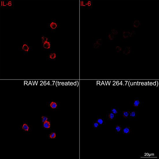
Confocal imaging of RAW 264.7 cells (treated with LPS and BFA) and RAW 264.7 cells (untreated) using IL-6 Rabbit PAb ( dilution 1:200) followed by a further incubation with Cy3-conjugated Goat Anti-Rabbit IgG (H+L) (AS007, dilution 1:500) (Red). DAPI was used for nuclear staining (Blue). Objective: 100x.

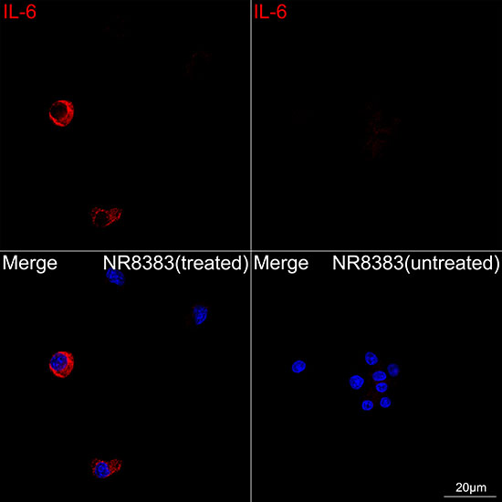
Confocal imaging of NR8383 cells (treated with LPS and BFA) and NR8383 cells (untreated) using IL-6 Rabbit pAb , dilution 1:200) followed by a further incubation with Cy3-conjugated Goat Anti-Rabbit IgG (H+L) (, dilution 1:500) (Red). DAPI was used for nuclear staining (Blue). Objective: 100x.

No documents available.