IL-6 Rabbit Polyclonal Antibody

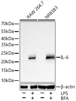
Western blot analysis of various lysates using IL-6 Rabbit PAb  at 1:5000 dilution incubated overnight at 4℃. RAW 264.7 cells and NR8383 cells were treated with LPS (1 μg/mL) and BFA (300 ng/mL) for 8 hours.
Secondary antibody: HRP-conjugated Goat anti-Rabbit IgG (H+L)  at 1:10000 dilution.
Lysates/proteins: 30 μg per lane.
Blocking buffer: 3% nonfat dry milk in TBST.
Detection: ECL West Pico Plus.
Exposure time: 45 s.

Western blot analysis of various lysates using IL-6 Rabbit PAb at 1:5000 dilution incubated overnight at 4℃. RAW 264.7 cells and NR8383 cells were treated with LPS (1 μg/mL) and BFA (300 ng/mL) for 8 hours. Secondary antibody: HRP-conjugated Goat anti-Rabbit IgG (H+L) at 1:10000 dilution. Lysates/proteins: 30 μg per lane. Blocking buffer: 3% nonfat dry milk in TBST. Detection: ECL West Pico Plus. Exposure time: 45 s.

Catalog Number:AB27935
Conjugate:Unconjugated
Size:100 ug
Concentration:1mg/ml
Host:Rabbit
Isotype:IgG
Immunogen:Recombinant protein. This information is considered to be commercially sensitive.
Reactivity:Mouse,Rat
Applications:WB 1:1000 - 1:5000 IP 0.5 μg-4 μg antibody for 900 μg-1100 μg extracts of whole cells IF/ICC 1:200 - 1:800 ELISA
Molecular Weight:24 kDa
Purification:Affinity purification
Form:liquid
Synonyms:ILg6; Ifnb2

Applications

WB 1:1000 - 1:5000 IP 0.5 μg-4 μg antibody for 900 μg-1100 μg extracts of whole cells IF/ICC 1:200 - 1:800 ELISA

Immunogen

Recombinant protein. This information is considered to be commercially sensitive.
Target Background

Enables cytokine activity and interleukin-6 receptor binding activity. Involved in several processes, including cellular response to cytokine stimulus; positive regulation of cell communication; and positive regulation of metabolic process. Located in extracellular space. Used to study several diseases, including acute necrotizing pancreatitis; disseminated intravascular coagulation; hypertension (multiple); impotence; and steatotic liver disease. Biomarker of several diseases, including acute necrotizing pancreatitis; artery disease (multiple); auditory system disease (multiple); gastrointestinal system cancer (multiple); and lung disease (multiple). Human ortholog(s) of this gene implicated in several diseases, including autoimmune disease (multiple); eye disease (multiple); gastrointestinal system cancer (multiple); glucose metabolism disease (multiple); and periodontal disease (multiple). Orthologous to human IL6 (interleukin 6).

Synonyms:ILg6; Ifnb2

Storage

Store at -20℃. Avoid freeze / thaw cycles.

Buffer

PBS containing 50% glycerol and 0.05% BSA, preserved with proclin300 or sodium azide (as specified on the Certificate of Analysis), pH 7.3.

Western blot analysis of various lysates using IL-6 Rabbit PAb at 1:5000 dilution incubated overnight at 4℃. RAW 264.7 cells and NR8383 cells were treated with LPS (1 μg/mL) and BFA (300 ng/mL) for 8 hours. Secondary antibody: HRP-conjugated Goat anti-Rabbit IgG (H+L) at 1:10000 dilution. Lysates/proteins: 30 μg per lane. Blocking buffer: 3% nonfat dry milk in TBST. Detection: ECL West Pico Plus. Exposure time: 45 s.

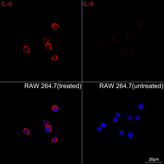
Confocal imaging of RAW 264.7 cells (treated with LPS and BFA) and RAW 264.7 cells (untreated) using IL-6 Rabbit PAb ( dilution 1:200) followed by a further incubation with Cy3-conjugated Goat Anti-Rabbit IgG (H+L) (AS007, dilution 1:500) (Red). DAPI was used for nuclear staining (Blue). Objective: 100x.

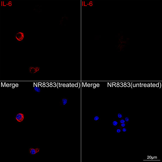
Confocal imaging of NR8383 cells (treated with LPS and BFA) and NR8383 cells (untreated) using IL-6 Rabbit pAb , dilution 1:200) followed by a further incubation with Cy3-conjugated Goat Anti-Rabbit IgG (H+L) (, dilution 1:500) (Red). DAPI was used for nuclear staining (Blue). Objective: 100x.

No documents available.