Interleukin-6

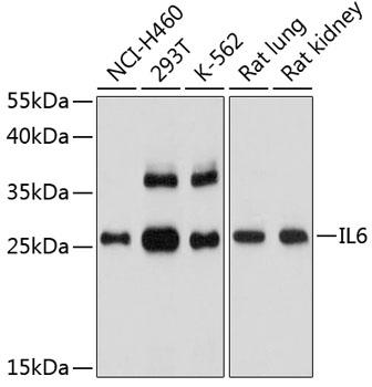
Western blot analysis of extracts of various 
cell lines, using IL6 antibody  at 1:1000 dilution.
Secondary antibody: HRP Goat Anti-Rabbit IgG 
(H+L) at 1:10000 dilution.
Lysates/proteins: 25ug per lane.
Blocking buffer: 3% nonfat dry milk in TBST.
Detection: ECL West Pico Plus.   
Exposure time: 90s.

Western blot analysis of extracts of various cell lines, using IL6 antibody at 1:1000 dilution. Secondary antibody: HRP Goat Anti-Rabbit IgG (H+L) at 1:10000 dilution. Lysates/proteins: 25ug per lane. Blocking buffer: 3% nonfat dry milk in TBST. Detection: ECL West Pico Plus. Exposure time: 90s.

Catalog Number:MAB-94635
Conjugate:Unconjugated
Size:100 ug
Concentration:1mg/ml
Host:Rabbit
Isotype:IgG
Clone:I621
Immunogen:The antiserum was produced against synthesized peptide derived from the Internal region of human IL6. AA range:131-180
Reactivity:Hu,Ms, Rt
Applications:Western Blot: 1/500 - 1/2000. Immunohistochemistry (paraffin-ebedded tissues): 1:100-300 Immunofluorescence: 1:100-300 ELISA: 1/20000
Molecular Weight:23kDa
Purification:The antibody was affinity-purified from rabbit antiserum by affinity-chromatography using epitope-specific immunogen.
Form:Liquid
Synonyms:IL6; IFNB2; Interleukin-6; IL-6; B-cell stimulatory factor 2; BSF-2; CTL differentiation factor; CDF; Hybridoma growth factor; Interferon beta-2; IFN-beta-2

Applications

Western Blot: 1/500 - 1/2000. Immunohistochemistry (paraffin-ebedded tissues): 1:100-300 Immunofluorescence: 1:100-300 ELISA: 1/20000

Immunogen

The antiserum was produced against synthesized peptide derived from the Internal region of human IL6. AA range:131-180

Target Background

This gene encodes a cytokine that functions in inflammation and the maturation of B cells. In addition, the encoded protein has been shown to be an endogenous pyrogen capable of inducing fever in people with autoimmune diseases or infections. The protein is primarily produced at sites of acute and chronic inflammation, where it is secreted into the serum and induces a transcriptional inflammatory response through interleukin 6 receptor, alpha. The functioning of this gene is implicated in a wide variety of inflammation-associated disease states, including suspectibility to diabetes mellitus and systemic juvenile rheumatoid arthritis. Alternative splicing results in multiple transcript variants.

Synonyms:IL6; IFNB2; Interleukin-6; IL-6; B-cell stimulatory factor 2; BSF-2; CTL differentiation factor; CDF; Hybridoma growth factor; Interferon beta-2; IFN-beta-2

Storage

Store at -20℃. Avoid freeze / thaw cycles.

Buffer

Liquid in PBS containing 50% glycerol, 0.5% BSA and 0.02% sodium azide.

Western blot analysis of extracts of various cell lines, using IL6 antibody at 1:1000 dilution. Secondary antibody: HRP Goat Anti-Rabbit IgG (H+L) at 1:10000 dilution. Lysates/proteins: 25ug per lane. Blocking buffer: 3% nonfat dry milk in TBST. Detection: ECL West Pico Plus. Exposure time: 90s.

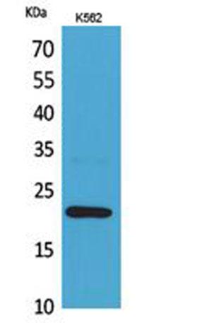
Western Blot analysis of K562 cells using IL-6 Antibody. Antibody was diluted at 1:1000. Secondary antibody was diluted at 1:20000

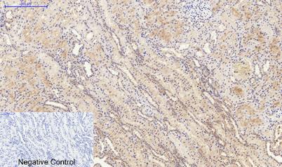
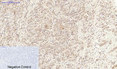
Immunohistochemical analysis of paraffin-embedded Human-breast-cancer tissue. 1,IL-6 Antibody was diluted at 1:200(4°C,overnight). 2, Sodium citrate pH 6.0 was used for antibody retrieval(>98°C,20min). 3,Secondary antibody was diluted at 1:200(room tempeRature, 30min). Negative control was used by secondary antibody only.

Immunohistochemical analysis of paraffin-embedded Human-kidney tissue. 1,IL-6 Antibody was diluted at 1:200(4°C,overnight). 2, Sodium citrate pH 6.0 was used for antibody retrieval(>98°C,20min). 3,Secondary antibody was diluted at 1:200(

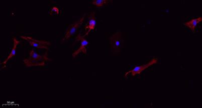
Immunofluorescence analysis of A549. 1,primary Antibody(red) was diluted at 1:200(4°C overnight). 2, Goat Anti Rabbit IgG (H&L) - Alexa Fluor 594 Secondary antibody was diluted at 1:1000(room temperature, 50min). 3, Picture B: DAPI(blue) 10min.

No documents available.