IRE1 Rabbit Monoclonal Antibody

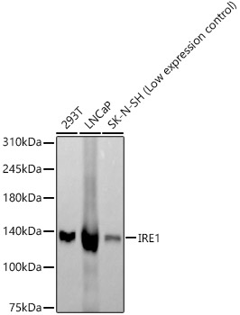
Western blot analysis of various lysates, using IRE1 antibody  at 1:1000 dilution.
Secondary antibody: HRP Goat Anti-Rabbit IgG (H+L)  at 1:10000 dilution.
Lysates/proteins: 25μg per lane. 
Blocking buffer: 3% nonfat dry milk in TBST. 
Detection: ECL West Pico Plus. 
Exposure time: 60s.

Western blot analysis of various lysates, using IRE1 antibody at 1:1000 dilution. Secondary antibody: HRP Goat Anti-Rabbit IgG (H+L) at 1:10000 dilution. Lysates/proteins: 25μg per lane. Blocking buffer: 3% nonfat dry milk in TBST. Detection: ECL West Pico Plus. Exposure time: 60s.

Catalog Number:MAB-94780
Conjugate:Unconjugated
Size:100 ug
Concentration:1mg/ml
Host:Rabbit
Isotype:IgG
Clone:ARC57190
Immunogen:Recombinant fusion protein containing a sequence corresponding to amino acids 286-435 of human IRE1.
Reactivity:Human, Mouse, Rat
Applications:Western Blot: 1:500-1:1000
Molecular Weight:130kDa
Purification:Affinity purification
Form:Liquid
Synonyms:IRE1; IRE1P; IRE1a; hIRE1p

Applications

Western Blot: 1:500-1:1000

Immunogen

Recombinant fusion protein containing a sequence corresponding to amino acids 286-435 of human IRE1.

Target Background

This gene encodes the transmembrane protein kinase inositol-requiring enzyme 1. The encoded protein contains two functional catalytic domains, a serine/threonine-protein kinase domain and an endoribonuclease domain. This protein functions as a sensor of unfolded proteins in the endoplasmic reticulum (ER) and triggers an intracellular signaling pathway termed the unfolded protein response (UPR). The UPR is an ER stress response that is conserved from yeast to mammals and activates genes involved in degrading misfolded proteins, regulating protein synthesis and activating molecular chaperones. This protein specifically mediates the splicing and activation of the stress response transcription factor X-box binding protein 1.

Synonyms:IRE1; IRE1P; IRE1a; hIRE1p

Storage

Store at -20℃. Avoid freeze / thaw cycles.

Buffer

PBS with 0.05% proclin300,0.05% BSA,50% glycerol,pH7.3.

Western blot analysis of various lysates, using IRE1 antibody at 1:1000 dilution. Secondary antibody: HRP Goat Anti-Rabbit IgG (H+L) at 1:10000 dilution. Lysates/proteins: 25μg per lane. Blocking buffer: 3% nonfat dry milk in TBST. Detection: ECL West Pico Plus. Exposure time: 60s.

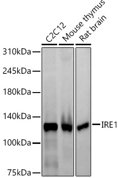
Western blot analysis of various lysates, using IRE1 antibody at 1:1000 dilution. Secondary antibody: HRP Goat Anti-Rabbit IgG (H+L) at 1:10000 dilution. Lysates/proteins: 25μg per lane. Blocking buffer: 3% nonfat dry milk in TBST. Detection: ECL West Pico Plus Exposure time: 120s.

No documents available.