
IRF7

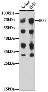
Western blot analysis of extracts of
various cell lines, using IRF7 antibody
at 1:1000 dilution.
Secondary antibody: HRP Goat Anti-
Rabbit IgG (H+L) at 1:10000
dilution.
Lysates/proteins: 25ug per lane.
Blocking buffer: 3% nonfat dry milk in
TBST.
Detection: ECL West Pico Plus.      
Exposure time: 15s.

Western blot analysis of extracts of various cell lines, using IRF7 antibody at 1:1000 dilution. Secondary antibody: HRP Goat Anti- Rabbit IgG (H+L) at 1:10000 dilution. Lysates/proteins: 25ug per lane. Blocking buffer: 3% nonfat dry milk in TBST. Detection: ECL West Pico Plus. Exposure time: 15s.

Catalog Number:MAB-94566
Conjugate:Unconjugated
Size:100ug
Concentration:1mg/ml
Host:Rb
Isotype:IgG
Clone:IF7
Immunogen:Recombinant fusion protein containing a sequence corresponding to amino acids 257-516 of human IRF7
Reactivity:Hu, Ms
Applications:Western Blot: 1:500 - 1:2000 Immunofluorescence: 1:50 - 1:200
Molecular Weight:70kDa
Purification:Aff. Pur.
Form:Liquid
Synonyms:IRF7;IMD39;IRF-7H;IRF7A;IRF7B;IRF7C;IRF7H

Applications

Western Blot: 1:500 - 1:2000 Immunofluorescence: 1:50 - 1:200

Immunogen

Recombinant fusion protein containing a sequence corresponding to amino acids 257-516 of human IRF7

Target Background

IRF7 encodes interferon regulatory factor 7, a member of the interferon regulatory transcription factor (IRF) family. IRF7 has been shown to play a role in the transcriptional activation of virus-inducible cellular genes, including interferon beta chain genes. Inducible expression of IRF7 is largely restricted to lymphoid tissue. Multiple IRF7 transcript variants have been identified, although the functional consequences of these have not yet been established.

Synonyms:IRF7;IMD39;IRF-7H;IRF7A;IRF7B;IRF7C;IRF7H

Storage

Store at -20℃. Avoid freeze / thaw cycles.

Buffer

PBS with 0.02% sodium azide,50% glycerol,pH7.3.

Western blot analysis of extracts of various cell lines, using IRF7 antibody at 1:1000 dilution. Secondary antibody: HRP Goat Anti- Rabbit IgG (H+L) at 1:10000 dilution. Lysates/proteins: 25ug per lane. Blocking buffer: 3% nonfat dry milk in TBST. Detection: ECL West Pico Plus. Exposure time: 15s.

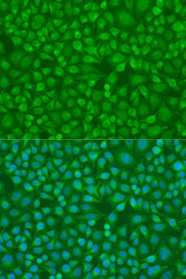
Immunofluorescence analysis of U2OS cells using IRF7 antibody at dilution of 1:100. Blue: DAPI for nuclear staining.

No documents available.