
IRS-1
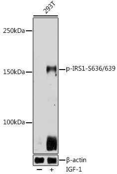
Western blot analysis of lysates from 293T cells, using Phospho-IRS1-S636/639 Rabbit pAb at 1:500 dilution. 293T cells were treated by IGF-1 (50 ng/ml) at 37℃ for 5 minutes after serum-starvation overnight. Secondary antibody: HRP-conjugated Goat anti-Rabbit IgG (H+L) at 1:10000 dilution. Lysates/proteins: 25μg per lane. Blocking buffer: 3% nonfat dry milk in TBST. Detection: ECL West Pico Plus. Exposure time: 10s.
| Catalog Number: | ABP0551 |
| Conjugate: | Unconjugated |
| Size: | 100ug |
| Concentration: | 1mg/ml |
| Host: | Rb |
| Isotype: | IgG |
| Clone: | POLY |
| Immunogen: | A synthetic phosphorylated peptide around S636 & S639 of human IRS1 (NP_005535.1) |
| Reactivity: | Human |
| Applications: | WB |
| Molecular Weight: | 180 kDa |
| Purification: | Affinity Purified |
| Form: | Liquid |
| Synonyms: | HIRS-1; Phospho-IRS1-S636/639 |
This gene encodes a protein which is phosphorylated by insulin receptor tyrosine kinase. Mutations in this gene are associated with type II diabetes and susceptibility to insulin resistance. RRID AB_2771218 Gene ID 3667 Swiss Prot Synonym HIRS-1; Phospho-IRS1-S636/639
Applications
WB
Immunogen
A synthetic phosphorylated peptide around S636 & S639 of human IRS1 (NP_005535.1)