JNK Rabbit Polyclonal Antibody

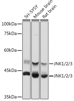
Western blot analysis of extracts of various cell lines, using JNK1/2/3 antibody  at 1:1000 dilution.
Secondary antibody: HRP Goat Anti-Rabbit IgG (H+L) at 1:10000 dilution.
Lysates/proteins: 25ug per lane.
Blocking buffer: 3% nonfat dry milk in TBST.
Detection: ECL West Pico Plus.
Exposure time: 5s.

Western blot analysis of extracts of various cell lines, using JNK1/2/3 antibody  at 1:1000 dilution. Secondary antibody: HRP Goat Anti-Rabbit IgG (H+L) at 1:10000 dilution. Lysates/proteins: 25ug per lane. Blocking buffer: 3% nonfat dry milk in TBST. Detection: ECL West Pico Plus. Exposure time: 5s.

Catalog Number:AB-82391
Conjugate:Unconjugated
Size:100 ug
Concentration:1mg/ml
Host:Rabbit
Isotype:IgG
Clone:POLY
Immunogen:Recombinant fusion protein containing a sequence corresponding to amino acids 1-370 of human JNK1/2/3.
Reactivity:Hu, Ms, Rt
Applications:Western Blot:                        1:500 - 1:2000 Immunohistochemistry:     1:50 - 1:200 Immunofluorescence:         1:50 - 1:200        Immunocytochemistry:      1:50 - 1:200 Immunoprecipitation:        1:500 - 1:1000
Molecular Weight:46kDa/54kDa
Purification:Aff. Pur.
Form:Liquid

Applications

Western Blot:                        1:500 - 1:2000 Immunohistochemistry:     1:50 - 1:200 Immunofluorescence:         1:50 - 1:200        Immunocytochemistry:      1:50 - 1:200 Immunoprecipitation:        1:500 - 1:1000

Immunogen

Recombinant fusion protein containing a sequence corresponding to amino acids 1-370 of human JNK1/2/3.

Target Background

JNK belongs to MAP kinase family. MAP kinases act as an integration point for multiple biochemical signals, and are involved in a wide variety of cellular processes such as proliferation, differentiation, transcription regulation and development. JNK is activated by various cell stimuli, and targets specific transcription factors, and thus mediates immediate-early gene expression in response to cell stimuli. The activation of this kinase by tumor-necrosis factor alpha (TNF-alpha) is found to be required for TNF-alpha induced apoptosis. This kinase is also involved in UV radiation induced apoptosis, which is thought to be related to cytochrom c-mediated cell death pathway. Studies of the mouse counterpart of this gene suggested that this kinase play a key role in T cell proliferation, apoptosis and differentiation.JNK is expressed as ten different isoforms due to differential mRNA splicing. The predominant forms are JNK1,JNK2,and JNK3.

Storage

Store at -20℃. Avoid freeze / thaw cycles.

Buffer

PBS with 0.05% proclin300,50% glycerol,pH7.3.

Western blot analysis of extracts of various cell lines, using JNK1/2/3 antibody  at 1:1000 dilution. Secondary antibody: HRP Goat Anti-Rabbit IgG (H+L) at 1:10000 dilution. Lysates/proteins: 25ug per lane. Blocking buffer: 3% nonfat dry milk in TBST. Detection: ECL West Pico Plus. Exposure time: 5s.

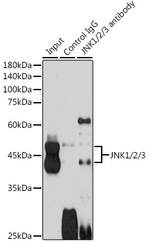
Immunoprecipitation analysis of 600ug extracts of Mouse brain cells using 3ug JNK1/2/3 antibody.                                                                       Western blot was performed from the immunoprecipitate using JNK1/2/3 antibody at a dilution of 1:1000.

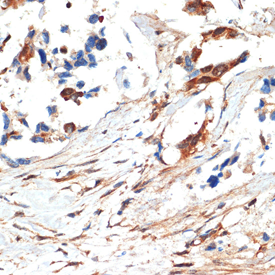
Immunohistochemistry of paraffinembedded human lung cancer using JNK1/2/3 Rabbit pAb at dilution of 1:100 (40x lens).Perform microwave antigen retrieval with 10 mM Tris/EDTA buffer pH 9.0 before commencing with IHC staining protocol.

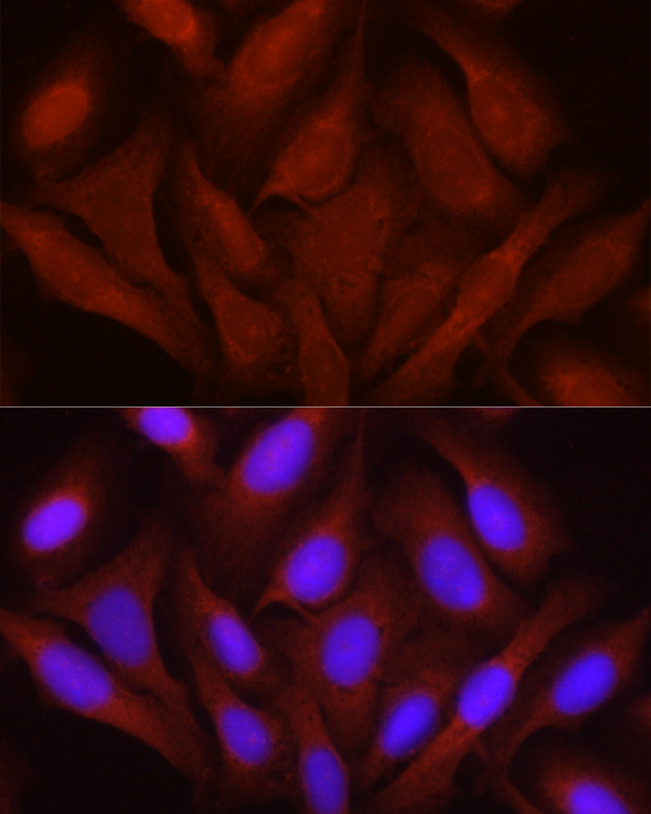
Immunofluorescence analysis of U2OS cells using JNK1/2/3 Rabbit pAb at dilution of 1:100 (40x lens).                                                                    Blue: DAPI for nuclear staining.

No documents available.