K-ras

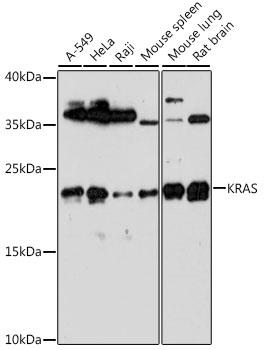
Western blot analysis of extracts of various cell lines, using KRAS antibody at 1:1000 dilution.
Secondary antibody: HRP Goat Anti-Rabbit IgG (H+L) at 1:10000 dilution.
Lysates/proteins: 25ug per lane.
Blocking buffer: 3% nonfat dry milk in TBST.
Detection: ECL West Pico.  
Exposure time: 90s.

Western blot analysis of extracts of various cell lines, using KRAS antibody at 1:1000 dilution. Secondary antibody: HRP Goat Anti-Rabbit IgG (H+L) at 1:10000 dilution. Lysates/proteins: 25ug per lane. Blocking buffer: 3% nonfat dry milk in TBST. Detection: ECL West Pico. Exposure time: 90s.

Catalog Number:MAB-94415
Size:100 ug
Concentration:1mg/ml
Host:Rb
Isotype:IgG
Clone:POLY
Immunogen:Recombinant protein of human KRAS
Reactivity:Hu, Ms, Rt
Applications:Western Blot: 1:500 -1:1000 Immunohistochemistry: 1:50 -1:200 Immunofluorescence: 1:50 -1:200 Immunoprecipitation: 1:50 -1:200
Molecular Weight:22kDa
Purification:Aff. Pur.
Form:Liquid
Synonyms:KRAS; C-K-RAS; CFC2; K-RAS2A; K-RAS2B; K-RAS4A; K-RAS4B; KI-RAS; KRAS1; KRAS2; NS; NS3; RALD; RASK2; c-Ki-ras2; GTPase Kras

Applications

Western Blot: 1:500 -1:1000 Immunohistochemistry: 1:50 -1:200 Immunofluorescence: 1:50 -1:200 Immunoprecipitation: 1:50 -1:200

Immunogen

Recombinant protein of human KRAS
Target Background

This gene, a Kirsten ras oncogene homolog from the mammalian ras gene family, encodes a protein that is a member of the small GTPase superfamily. A single amino acid substitution is responsible for an activating mutation. The transforming protein that results is implicated in various malignancies, including lung adenocarcinoma, mucinous adenoma, ductal carcinoma of the pancreas and colorectal carcinoma. Alternative splicing leads to variants encoding two isoforms that differ in the C-terminal region.

Synonyms:KRAS; C-K-RAS; CFC2; K-RAS2A; K-RAS2B; K-RAS4A; K-RAS4B; KI-RAS; KRAS1; KRAS2; NS; NS3; RALD; RASK2; c-Ki-ras2; GTPase Kras

Storage

Store at -20℃. Avoid freeze / thaw cycles

Buffer

PBS with 0.02% sodium azide, 50% glycerol, pH7.3.

Western blot analysis of extracts of various cell lines, using KRAS antibody at 1:1000 dilution. Secondary antibody: HRP Goat Anti-Rabbit IgG (H+L) at 1:10000 dilution. Lysates/proteins: 25ug per lane. Blocking buffer: 3% nonfat dry milk in TBST. Detection: ECL West Pico. Exposure time: 90s.

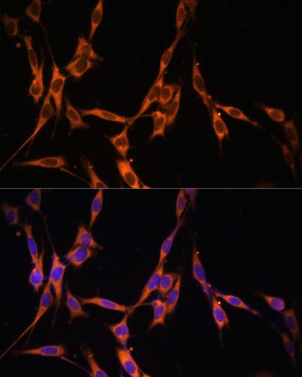
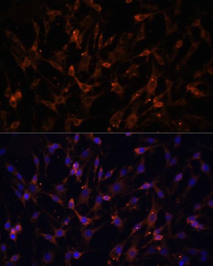
Immunofluorescence analysis of C6 cells using KRAS antibody at dilution of 1:100. Blue: DAPI for nuclear staining.

Immunofluorescence analysis of NIH/3T3 cells using KRAS antibody at dilution of 1:100. Blue: DAPI for nuclear staining.

No documents available.