[KD Validated] COX5A Rabbit mAb

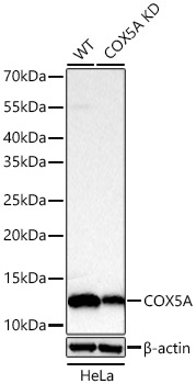
Western blot analysis of lysates from wild type (WT) and COX5A knockdown (KD) HeLa cells using [KD Validated] COX5A Rabbit mAb (A25751) at 1:2000 dilution incubated overnight at 4℃.
Secondary antibody: HRP-conjugated Goat anti-Rabbit IgG (H+L) (AS014) at 1:10000 dilution.
Lysates/proteins: 25 μg per lane.
Blocking buffer: 3% nonfat dry milk in TBST.
Detection: ECL Basic Kit (RM00020)
Exposure time: 30 s.

Western blot analysis of lysates from wild type (WT) and COX5A knockdown (KD) HeLa cells using [KD Validated] COX5A Rabbit mAb (A25751) at 1:2000 dilution incubated overnight at 4℃. Secondary antibody: HRP-conjugated Goat anti-Rabbit IgG (H+L) (AS014) at 1:10000 dilution. Lysates/proteins: 25 μg per lane. Blocking buffer: 3% nonfat dry milk in TBST. Detection: ECL Basic Kit (RM00020) Exposure time: 30 s.

Catalog Number:AB25751
Conjugate:Unconjugated
Size:100 ug
Concentration:1mg/ml
Host:Rabbit
Isotype:IgG
Clone:ARC67578
Immunogen:Recombinant fusion protein containing a sequence corresponding to amino acids 1-150 of human COX5A.
Reactivity:Human,Mouse,Rat
Applications:WB 1:2000 - 1:8000 IHC-P 1:300 - 1:3000 IF/ICC 1:200 - 1:800 ELISA Recommended starting concentration is 1 μg/mL. Please optimize the concentration based on your specific assay requirements.
Molecular Weight:13kDa
Purification:Affinity purification
Form:liquid
Synonyms:VA; COX; COX-VA; MC4DN20

Cytochrome c oxidase (COX) is the terminal enzyme of the mitochondrial respiratory chain. It is a multi-subunit enzyme complex that couples the transfer of electrons from cytochrome c to molecular oxygen and contributes to a proton electrochemical gradient across the inner mitochondrial membrane. The complex consists of 13 mitochondrial- and nuclear-encoded subunits. The mitochondrially-encoded subunits perform the electron transfer of proton pumping activities. The functions of the nuclear-encoded subunits are unknown but they may play a role in the regulation and assembly of the complex. This gene encodes the nuclear-encoded subunit Va of the human mitochondrial respiratory chain enzyme. A pseudogene COX5AP1 has been found in chromosome 14q22.

Applications

WB 1:2000 - 1:8000 IHC-P 1:300 - 1:3000 IF/ICC 1:200 - 1:800 ELISA Recommended starting concentration is 1 μg/mL. Please optimize the concentration based on your specific assay requirements.

Immunogen

Recombinant fusion protein containing a sequence corresponding to amino acids 1-150 of human COX5A.

Target Background

Cytochrome c oxidase (COX) is the terminal enzyme of the mitochondrial respiratory chain. It is a multi-subunit enzyme complex that couples the transfer of electrons from cytochrome c to molecular oxygen and contributes to a proton electrochemical gradient across the inner mitochondrial membrane. The complex consists of 13 mitochondrial- and nuclear-encoded subunits. The mitochondrially-encoded subunits perform the electron transfer of proton pumping activities. The functions of the nuclear-encoded subunits are unknown but they may play a role in the regulation and assembly of the complex. This gene encodes the nuclear-encoded subunit Va of the human mitochondrial respiratory chain enzyme. A pseudogene COX5AP1 has been found in chromosome 14q22.

Synonyms:VA; COX; COX-VA; MC4DN20

Storage

Store at -20℃. Avoid freeze / thaw cycles.

Buffer

PBS with 0.05% proclin300,0.05% BSA,50% glycerol,pH7.3.

Western blot analysis of lysates from wild type (WT) and COX5A knockdown (KD) HeLa cells using [KD Validated] COX5A Rabbit mAb (A25751) at 1:2000 dilution incubated overnight at 4℃. Secondary antibody: HRP-conjugated Goat anti-Rabbit IgG (H+L) (AS014) at 1:10000 dilution. Lysates/proteins: 25 μg per lane. Blocking buffer: 3% nonfat dry milk in TBST. Detection: ECL Basic Kit (RM00020) Exposure time: 30 s.

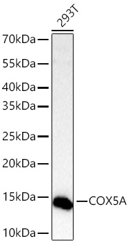
Western blot analysis of lysates from 293T cells using [KD Validated] COX5A Rabbit mAb (A25751) at 1:2000 dilution incubated overnight at 4℃. Secondary antibody: HRP-conjugated Goat anti-Rabbit IgG (H+L) (AS014) at 1:10000 dilution. Lysates/proteins: 25 μg per lane. Blocking buffer: 3% nonfat dry milk in TBST. Detection: ECL Basic Kit (RM00020) Exposure time: 30 s.

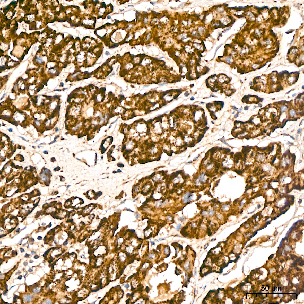
Immunohistochemistry analysis of paraffin-embedded Human liver tissue using [KD Validated] COX5A Rabbit mAb (A25751) at a dilution of 1:300 (40x lens). High pressure antigen retrieval performed with 0.01M Citrate Bufferr (pH 6.0) prior to IHC staining.

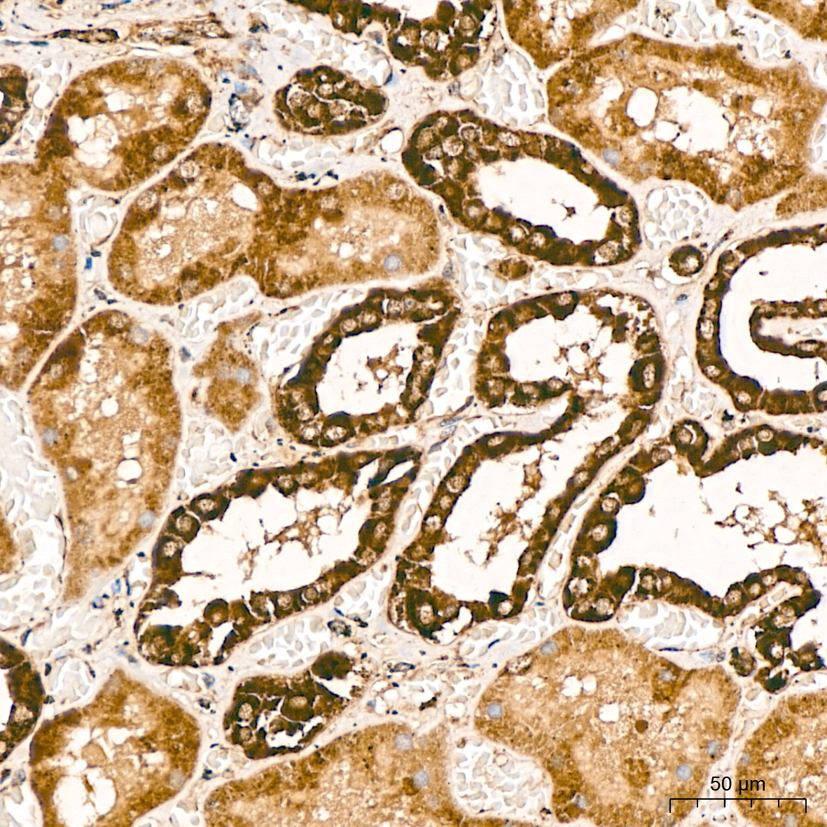
Immunohistochemistry analysis of paraffin-embedded Human kidney tissue using [KD Validated] COX5A Rabbit mAb (A25751) at a dilution of 1:300 (40x lens). High pressure antigen retrieval performed with 0.01M Citrate Bufferr (pH 6.0) prior to IHC staining.

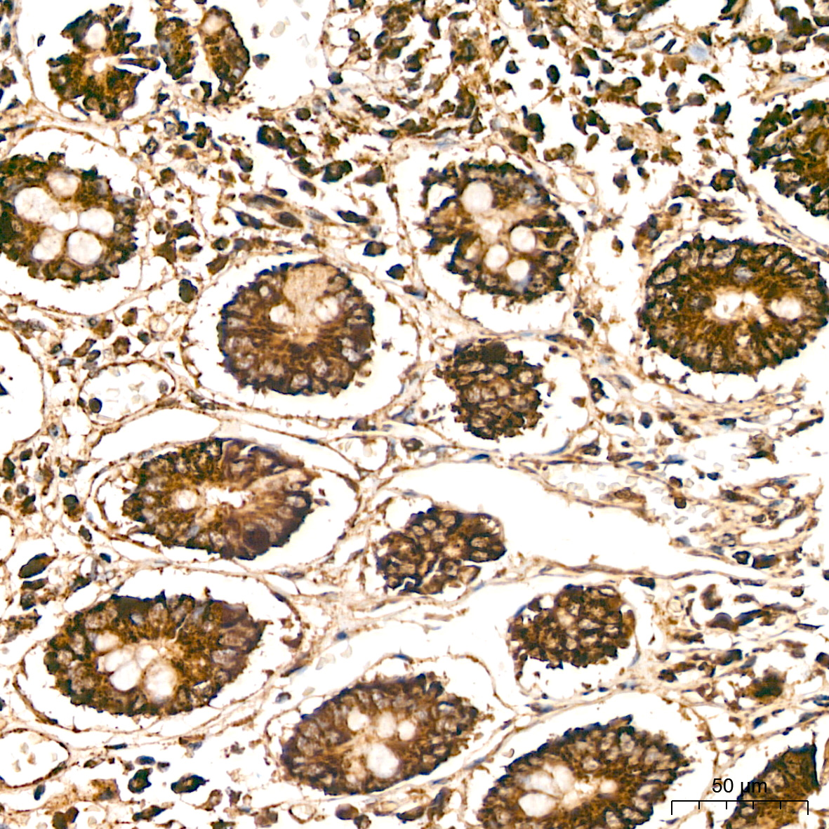
Immunohistochemistry analysis of paraffin-embedded Human colon tissue using [KD Validated] COX5A Rabbit mAb (A25751) at a dilution of 1:300 (40x lens). High pressure antigen retrieval performed with 0.01M Citrate Bufferr (pH 6.0) prior to IHC staining.

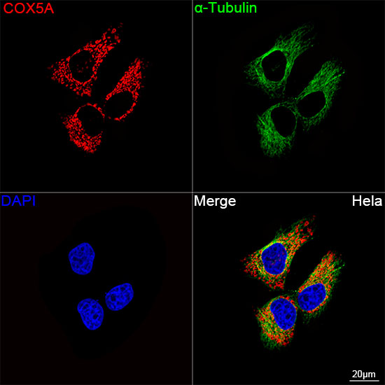
Confocal imaging of HeLa cells using [KD Validated] COX5A Rabbit mAb (A25751, dilution 1:200) followed by a further incubation with Cy3 Goat Anti-Rabbit IgG (H+L) (AS007, dilution 1:500) (Red). The cells were counterstained with α-Tubulin Mouse mAb (AC012, dilution 1:400) followed by incubation with ABflo® 488-conjugated Goat Anti-Mouse IgG (H+L) Ab (AS076, dilution 1:500) (Green). DAPI was used for nuclear staining (Blue). Objective: 100x.

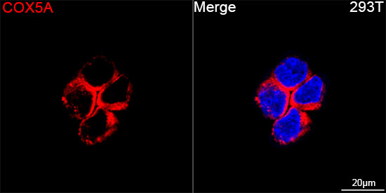
Confocal imaging of 293T cells using [KD Validated] COX5A Rabbit mAb (A25751, dilution 1:200) followed by a further incubation with Cy3 Goat Anti-Rabbit IgG (H+L) (AS007, dilution 1:500) (Red). DAPI was used for nuclear staining (Blue). Objective: 100x.

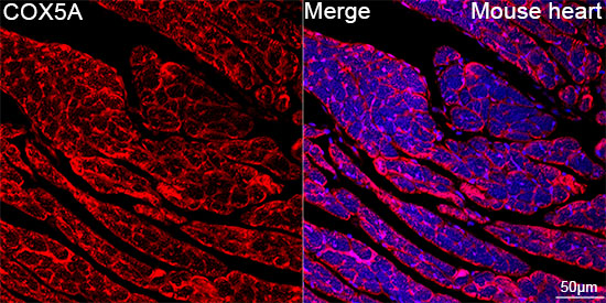
Confocal imaging of paraffin-embedded Mouse heart tissue using [KD Validated] COX5A Rabbit mAb (A25751, dilution 1:200) followed by a further incubation with Cy3 Goat Anti-Rabbit IgG (H+L) (AS007, dilution 1:500) (Red). DAPI was used for nuclear staining (Blue). High pressure antigen retrieval performed with 0.01M Citrate Buffer (pH 6.0) prior to IF staining. Objective: 40x.

Confocal imaging of paraffin-embedded Rat brain tissue using [KD Validated] COX5A Rabbit mAb (A25751, dilution 1:200) followed by a further incubation with Cy3 Goat Anti-Rabbit IgG (H+L) (AS007, dilution 1:500) (Red). DAPI was used for nuclear staining (Blue). Microwave antigen retrieval performed with 0.01M Citrate Buffer (pH 6.0) prior to IF staining. Objective: 40x.

No documents available.