
KEAP1

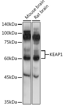
Western blot analysis of extracts of various cell lines, using KEAP1 antibody at 1:1000 dilution. 
Secondary antibody: HRP Goat Anti-Rabbit IgG (H+L)  at 1:10000 dilution. 
Lysates/proteins: 25ug per lane. 
Blocking buffer: 3% nonfat dry milk in TBST. Detection: ECL West Pico Plus.     
Exposure time: 60s.

Western blot analysis of extracts of various cell lines, using KEAP1 antibody at 1:1000 dilution. Secondary antibody: HRP Goat Anti-Rabbit IgG (H+L) at 1:10000 dilution. Lysates/proteins: 25ug per lane. Blocking buffer: 3% nonfat dry milk in TBST. Detection: ECL West Pico Plus. Exposure time: 60s.

Catalog Number:AB-83755
Conjugate:Unconjugated
Size:100 ug
Concentration:1mg/ml
Host:Rabbit
Isotype:IgG
Clone:POLY
Immunogen:A synthetic peptide corresponding to a sequence within amino acids 300-400 of human KEAP1.
Reactivity:Hu, Ms, Rt
Applications:Western Blot: 1:500 - 1:1000 Immunofluorescence: 1:50 - 1:200 Immunocytochemistry: 1:50 - 1:200
Molecular Weight:60-64 kDa
Purification:Aff. Pur.
Form:Liquid
Synonyms:KEAP1;INrf2;KLHL19

Applications

Western Blot: 1:500 - 1:1000 Immunofluorescence: 1:50 - 1:200 Immunocytochemistry: 1:50 - 1:200

Immunogen

A synthetic peptide corresponding to a sequence within amino acids 300-400 of human KEAP1.
Target Background

This gene encodes a protein containing KELCH-1 like domains, as well as a BTB/POZ domain. Kelch-like ECH-associated protein 1 interacts with NF-E2-related factor 2 in a redox-sensitive manner and the dissociation of the proteins in the cytoplasm is followed by transportation of NF-E2-related factor 2 to the nucleus. This interaction results in the expression of the catalytic subunit of gammaglutamylcysteine synthetase. Two alternatively spliced transcript variants encoding the same isoform have been found for this gene.

Synonyms:KEAP1;INrf2;KLHL19

Storage

Store at -20℃. Avoid freeze / thaw cycles.

Buffer

PBS with 0.01% thiomersal,50% glycerol,pH7.3.

Western blot analysis of extracts of various cell lines, using KEAP1 antibody at 1:1000 dilution. Secondary antibody: HRP Goat Anti-Rabbit IgG (H+L) at 1:10000 dilution. Lysates/proteins: 25ug per lane. Blocking buffer: 3% nonfat dry milk in TBST. Detection: ECL West Pico Plus. Exposure time: 60s.

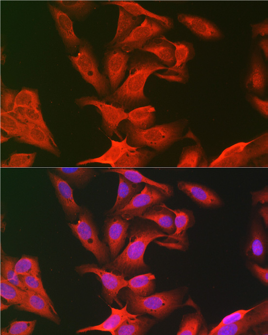
Immunofluorescence analysis of U2OS cells using KEAP1 Rabbit pAb at dilution of 1:100 (40x lens). Blue: DAPI for nuclear staining.

No documents available.