Ki-67

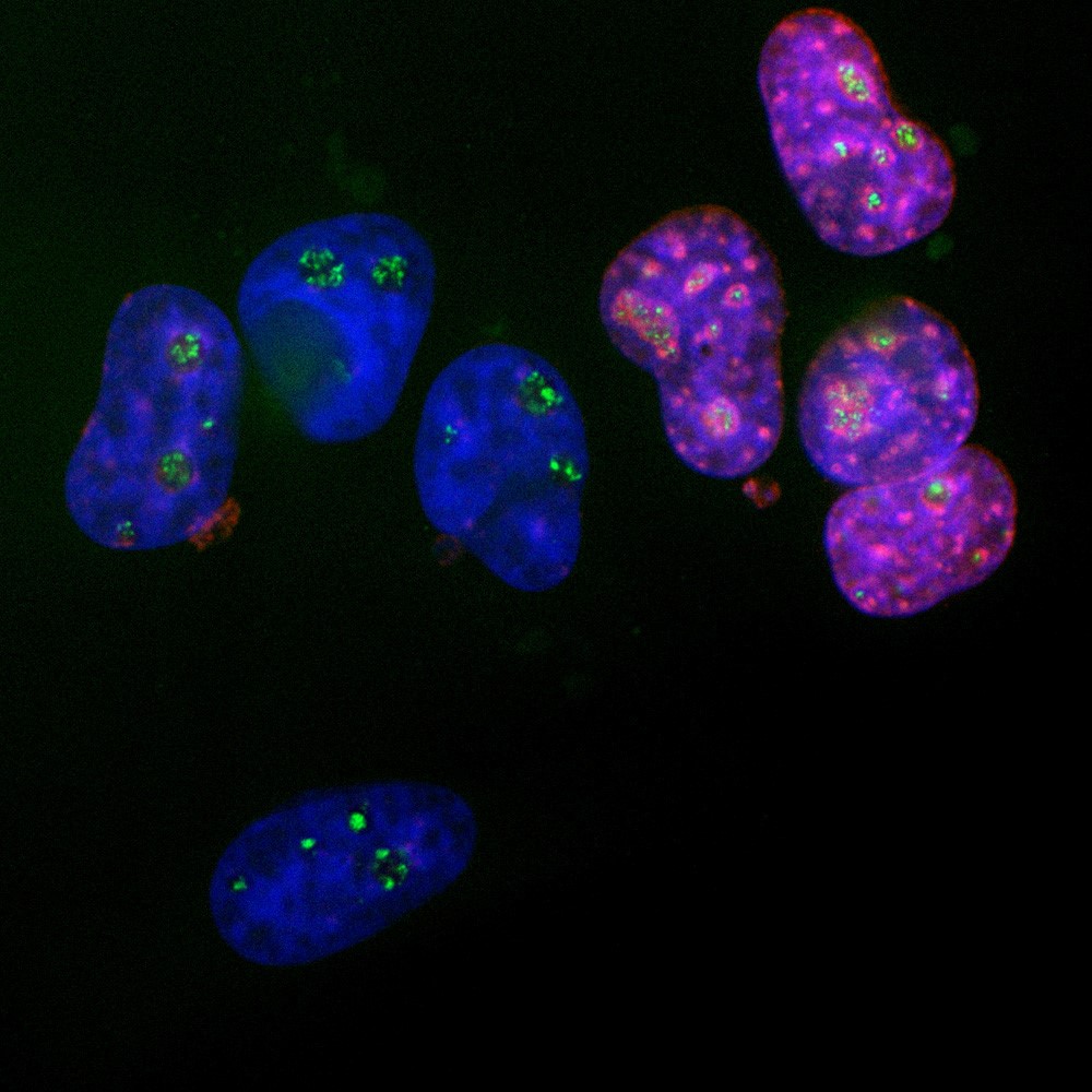
Immunofluorescent analysis of HeLa cells stained with rabbit Anti-Ki-67 dilution 1:2,500 in red, and mouse monoclonal antibody to Fibrillarin, dilution 1:2,000, in green. The blue is DAPI staining of nuclear DNA.

Immunofluorescent analysis of HeLa cells stained with rabbit Anti-Ki-67 dilution 1:2,500 in red, and mouse monoclonal antibody to Fibrillarin, dilution 1:2,000, in green. The blue is DAPI staining of nuclear DNA.

Catalog Number:AB-90972
Conjugate:Unconjugated
Size:100 ug
Concentration:1mg/ml
Host:Rb
Isotype:IgG
Clone:POLY
Immunogen:Recombinant human construct containing amino acids 1,111-1,490 expressed in and purified from E. coli.
Reactivity:Hu, Ms, Rt
Applications:Western Blot : 1:2,000-1:5,000 Immunohistochemsitry (paraffin, formalin, frozen Tissues): 1:500 Immunofluorescence/Immunocytochemistry: 1:1,000-1:2,500
Molecular Weight:345-395kDa
Purification:serum
Form:Liquid

Applications

Western Blot : 1:2,000-1:5,000 Immunohistochemsitry (paraffin, formalin, frozen Tissues): 1:500 Immunofluorescence/Immunocytochemistry: 1:1,000-1:2,500

Immunogen

Recombinant human construct containing amino acids 1,111-1,490 expressed in and purified from E. coli.

Target Background

The Ki-67 proteins were first discovered in an attempt to generate cancer specific monoclonal antibodies. A monoclonal antibody which bound to structures in the nuclei of dividing but not quiescent cells was produced and shown to bind two very large proteins of molecular weight 345kDa and 395kDa. The two proteins were derived from alternate transcripts of a single gene. The presence of Ki-67 proteins, detected with an appropriate antibody, is an indicator of cell proliferation and the level of Ki-67 expression is one of the most reliable biomarkers of proliferative status of cancer cells. The RPCA-Ki-67 antibody was raised against a recombinant construct containing amino acids 1,111-1,490 of human Ki-67 isotype 1. The antibody can be used to identify dividing cells in rat and mouse brain and also works on paraffin sections of human tissues, where it is useful to identify cancer cells. Mouse select image at left for larger view.

Storage

Storage for short term at 4°C recommended, for longer term at -20°C, minimize freeze/thaw cycles

Buffer

Supplied as an aliquot of serum plus 5mM sodium azide.

Immunofluorescent analysis of HeLa cells stained with rabbit Anti-Ki-67 dilution 1:2,500 in red, and mouse monoclonal antibody to Fibrillarin, dilution 1:2,000, in green. The blue is DAPI staining of nuclear DNA.

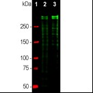
Western blot analysis of equal amounts of cell lysates using rabbit Anti-Ki-67 Ki-67, dilution 1:5,000, (green): [1] protein standard (red), [2] HeLa cells, [3] HEK293 cells. Strong double bands above 250kDa correspond to the two major Ki-67 isoforms of molecular weight 345kDa and 395kDa. Smaller proteolytic fragments of these isoforms are also detected on the blot.

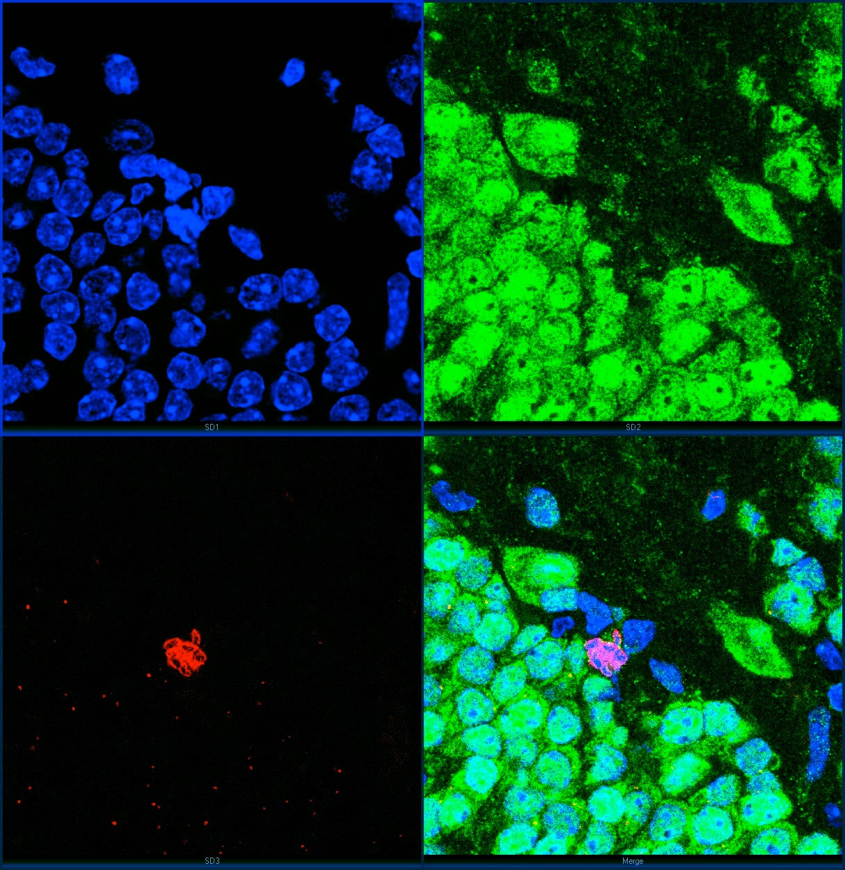
High magnification confocal image of adult mouse hippocampus dentate region stained with Anti-Ki67 Polyclonal 1:2,000 – in green. Blue is Hoechst dye staining of DNA. Top left is DNA, top right FOX3/NeuN, bottom left Ki-67 and bottom right all three merged. Dividing cells are very rare in adult animals, but one can be seen in the center of the image. Chromosomes can be seen in blue and their Ki-67 coating can be seen in red. The dividing cell is FOX3/NeuN negative and so is presumably a glial cell.

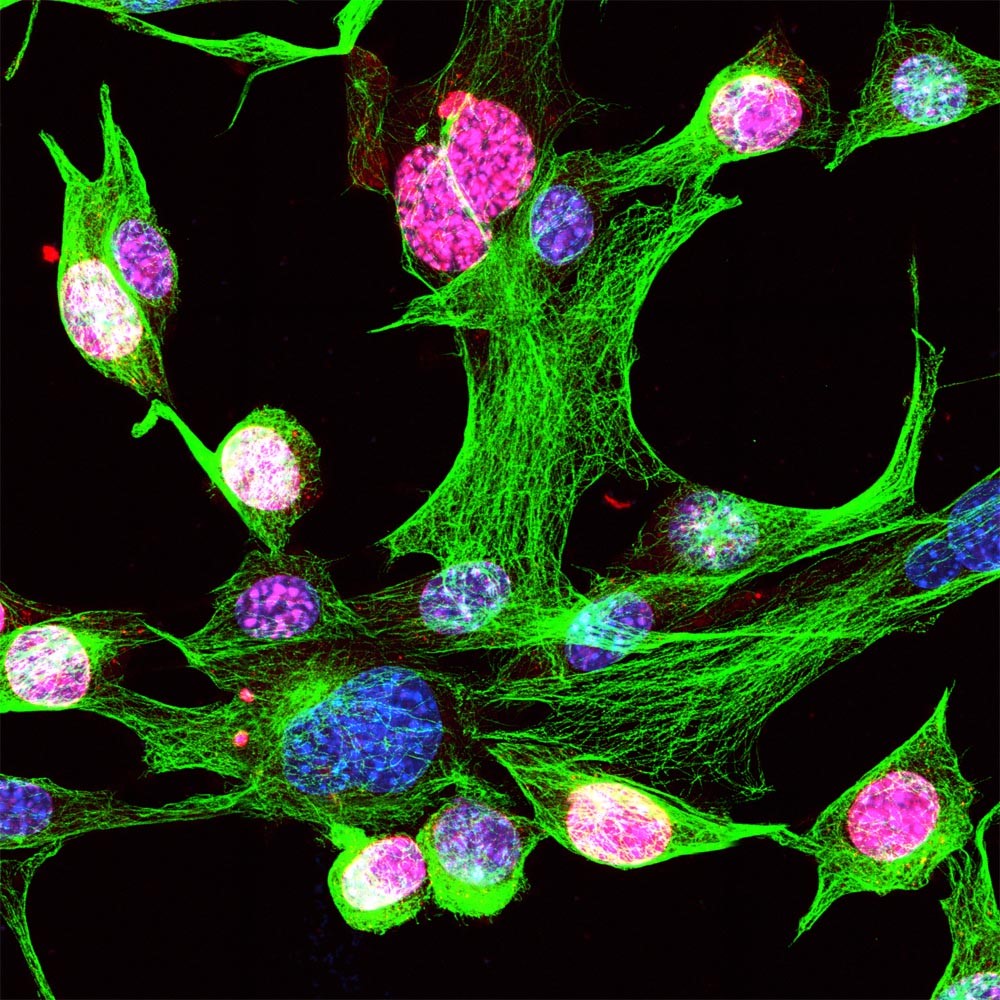
Mouse NIH-3T3 cells stained with Anti-Ki-67 1:2,500 in red and mouse mAb to β-tubulin, 1:1,000 in green. The Ki-67 strongly stains the nuclei of dividing cells, but not quiescent cells

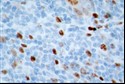
A section of human breast tissue including both normal and cancer cells. The cancer cells divide rapidly and heavily express Ki-67 and so stain strongly with the Anti-Ki-67 antibody.

No documents available.