
KI-67
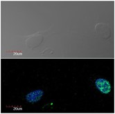
Immunocytochemistry detection of Ki-67 in U2OS cell line (human osteosarcoma) using monoclonal antibody Ki-67 (green). Cell nuclei stained with DAPI (blue)
| Catalog Number: | MAB-94467 |
| Conjugate: | Unconjugated |
| Size: | 100 ul |
| Concentration: | 1mg/ml |
| Host: | Ms |
| Isotype: | IgG1 |
| Clone: | Mib1 |
| Immunogen: | Nuclei of the Hodgkin lymphoma cell line L428 |
| Reactivity: | Hu |
| Applications: | Immunohistochemistry , Western Blot , Flow Cytometry (QC tested), Immunocytochemistry |
| Purification: | Aff. Pur. |
| Form: | Liquid |
| Synonyms: | KIA, MIB, Marker of proliferation Ki67, MKI67 |
Applications
Immunohistochemistry , Western Blot , Flow Cytometry (QC tested), Immunocytochemistry
Immunogen
Nuclei of the Hodgkin lymphoma cell line L428