[KO Validated] NF-kB p65/RelA Rabbit Monoclonal Antibody

Western blot analysis of lysates from wild type (WT) and NF-kB p65/RelA knockout (KO) HeLa cells using [KO Validated] NF-kB p65/RelA Rabbit mAb  at 1:10000 dilution incubated overnight at 4℃.
Secondary antibody: HRP-conjugated Goat anti-Rabbit IgG (H+L) at 1:10000 dilution.
Lysates/proteins: 25 μg per lane.
Blocking buffer: 3% nonfat dry milk in TBST.
Detection: ECL West Pico Plus.      
Exposure time: 30s.

Western blot analysis of lysates from wild type (WT) and NF-kB p65/RelA knockout (KO) HeLa cells using [KO Validated] NF-kB p65/RelA Rabbit mAb at 1:10000 dilution incubated overnight at 4℃. Secondary antibody: HRP-conjugated Goat anti-Rabbit IgG (H+L) at 1:10000 dilution. Lysates/proteins: 25 μg per lane. Blocking buffer: 3% nonfat dry milk in TBST. Detection: ECL West Pico Plus. Exposure time: 30s.

Catalog Number:MAB-94597
Conjugate:Unconjugated
Size:100 ug
Concentration:1mg/ml
Host:Rabbit
Isotype:IgG
Clone:ARC51086
Immunogen:Synthetic peptide. This information is considered to be commercially sensitive.
Reactivity:Human,Mouse,Rat,Monkey
Applications:WB 1:5000 - 1:20000 IHC-P 1:2000 - 1:8000 IF/ICC 1:600 - 1:2400 ChIP 5μg antibody for 10μg-15μg of Chromatin ELISA Recommended starting concentration is 1 μg/mL. Please optimize the concentration based on your specific assay requirements.
Molecular Weight:65kDa
Purification:Affinity purification
Form:liquid
Synonyms:p65; CMCU; NFKB3; AIF3BL3; NF-kB p65/RelA

NF-kappa-B is a ubiquitous transcription factor involved in several biological processes. It is held in the cytoplasm in an inactive state by specific inhibitors. Upon degradation of the inhibitor, NF-kappa-B moves to the nucleus and activates transcription of specific genes. NF-kappa-B is composed of NFKB1 or NFKB2 bound to either REL, RELA, or RELB. The most abundant form of NF-kappa-B is NFKB1 complexed with the product of this gene, RELA. Four transcript variants encoding different isoforms have been found for this gene.

Applications

WB 1:5000 - 1:20000 IHC-P 1:2000 - 1:8000 IF/ICC 1:600 - 1:2400 ChIP 5μg antibody for 10μg-15μg of Chromatin ELISA Recommended starting concentration is 1 μg/mL. Please optimize the concentration based on your specific assay requirements.

Immunogen

Synthetic peptide. This information is considered to be commercially sensitive.
Target Background

NF-kappa-B is a ubiquitous transcription factor involved in several biological processes. It is held in the cytoplasm in an inactive state by specific inhibitors. Upon degradation of the inhibitor, NF-kappa-B moves to the nucleus and activates transcription of specific genes. NF-kappa-B is composed of NFKB1 or NFKB2 bound to either REL, RELA, or RELB. The most abundant form of NF-kappa-B is NFKB1 complexed with the product of this gene, RELA. Four transcript variants encoding different isoforms have been found for this gene.

Synonyms:p65; CMCU; NFKB3; AIF3BL3; NF-kB p65/RelA

Storage

Store at -20℃. Avoid freeze / thaw cycles.

Buffer

PBS with 0.09% Sodium azide,0.05% BSA,50% glycerol,pH7.3.

Western blot analysis of lysates from wild type (WT) and NF-kB p65/RelA knockout (KO) HeLa cells using [KO Validated] NF-kB p65/RelA Rabbit mAb at 1:10000 dilution incubated overnight at 4℃. Secondary antibody: HRP-conjugated Goat anti-Rabbit IgG (H+L) at 1:10000 dilution. Lysates/proteins: 25 μg per lane. Blocking buffer: 3% nonfat dry milk in TBST. Detection: ECL West Pico Plus. Exposure time: 30s.

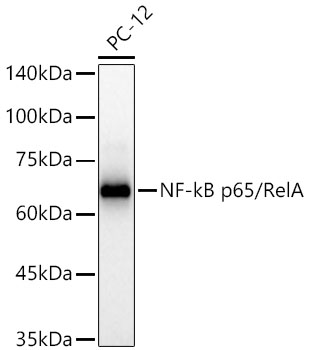
Western blot analysis of lysates from PC-12 cells using [KO Validated] NF-kB p65/RelA Rabbit mAb at 1:10000 dilution incubated overnight at 4℃. Secondary antibody: HRP-conjugated Goat anti-Rabbit IgG (H+L) at 1:10000 dilution. Lysates/proteins: 25 μg per lane. Blocking buffer: 3% nonfat dry milk in TBST. Detection: ECL West Pico Plus. Exposure time: 30s.

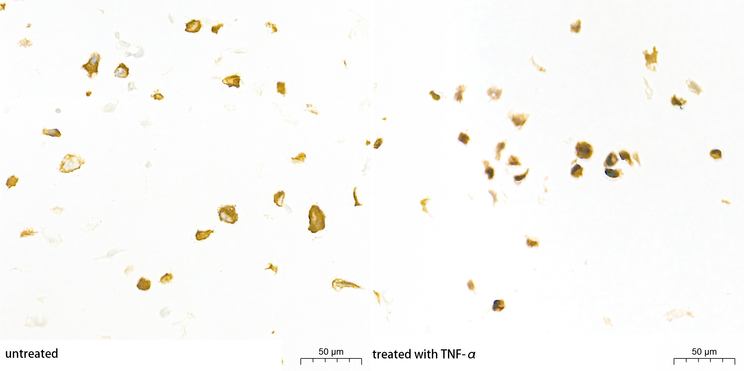
Immunohistochemistry analysis of paraffin-embedded HT-1080 cell lines(untreated and treated with TNF-α) using [KO Validated] NF-kB p65/RelA Rabbit mAb at a dilution of 1:3000 (40x lens). High pressure antigen retrieval performed with 0.01M Tris-EDTA Buffer (pH 9.0) prior to IHC staining.

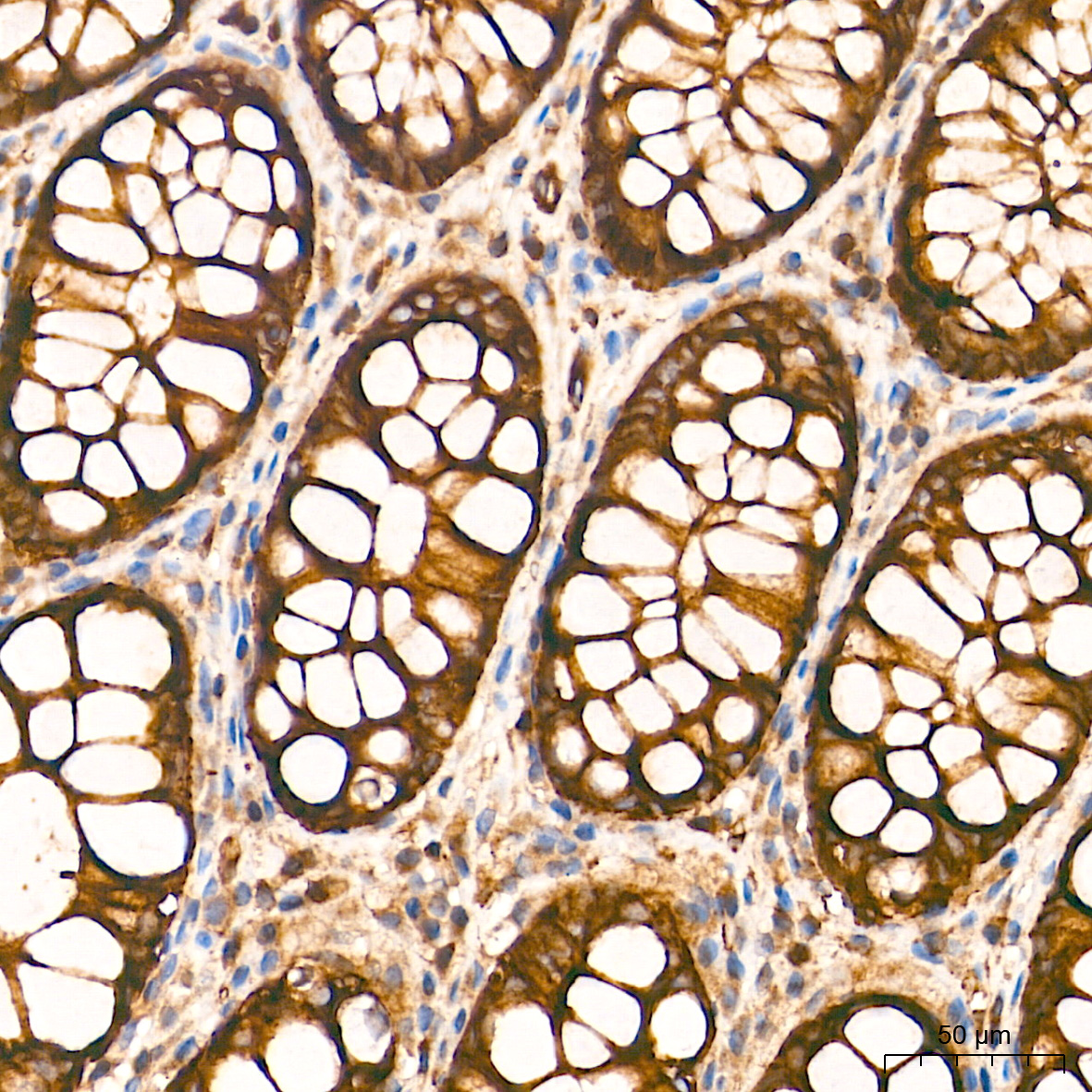
Immunohistochemistry analysis of paraffin-embedded Human colon tissue using [KO Validated] NF-kB p65/RelA Rabbit mAb at a dilution of 1:3000 (40x lens). High pressure antigen retrieval performed with 0.01M Tris-EDTA Buffer (pH 9.0) prior to IHC staining.

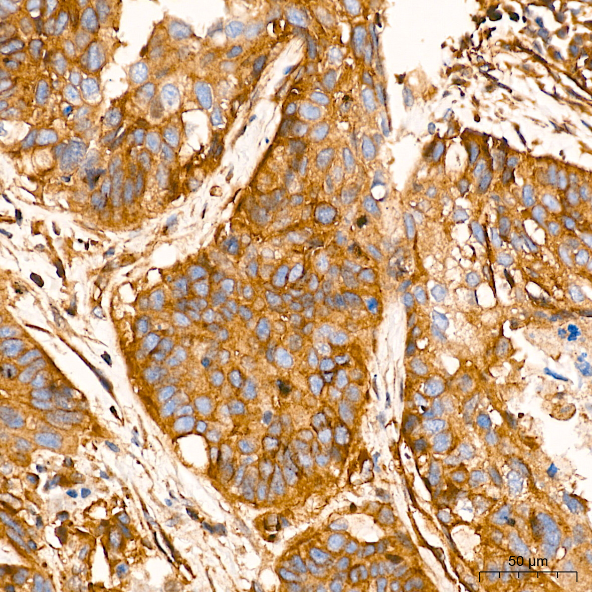
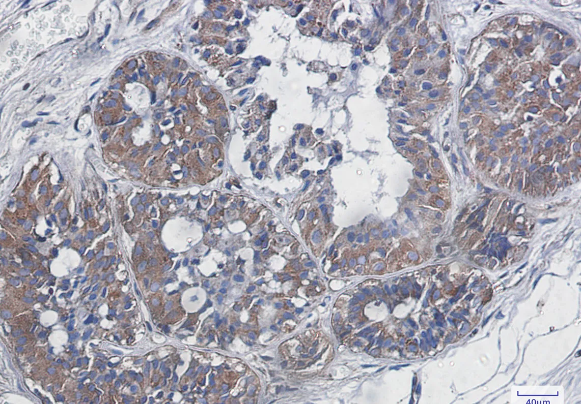
Immunohistochemistry analysis of paraffin-embedded Human lung cancer tissue using [KO Validated] NF-kB p65/RelA Rabbit mAb at a dilution of 1:3000 (40x lens). High pressure antigen retrieval performed with 0.01M Tris-EDTA Buffer (pH 9.0) prior to IHC staining.

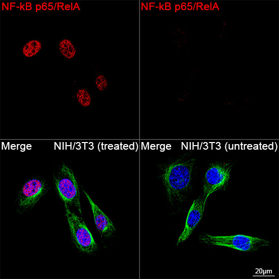
Confocal imaging of NIH/3T3 cells (treated with TNF-α) and NIH/3T3 cells (untreated) cells using [KO Validated] NF-kB p65/RelA Rabbit mAb (dilution 1:2100) followed by a further incubation with Cy3 Goat Anti-Rabbit IgG (H+L) (dilution 1:500) (Red). The cells were counterstained with α-Tubulin Mouse mAb (AC012, dilution 1:400) followed by incubation with ABflo® 488-conjugated Goat Anti-Mouse IgG (H+L) Ab , dilution 1:500) (Green). DAPI was used for nuclear staining (Blue). Objective: 100x.

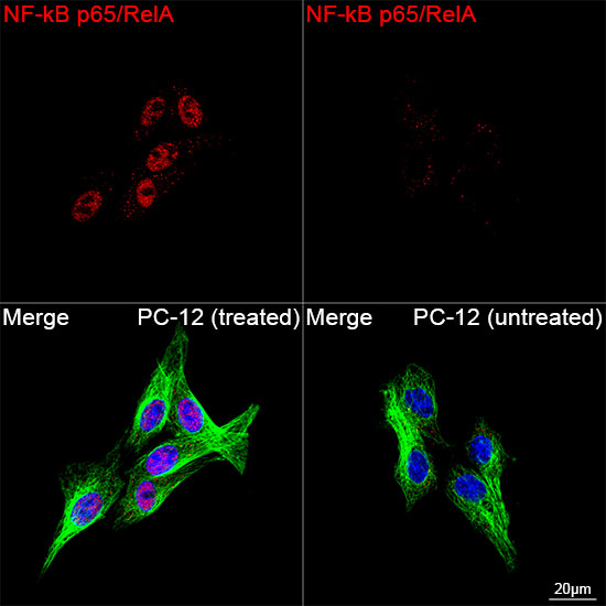
Confocal imaging of PC-12 cells (treated with TNF-α) and PC-12 cells (untreated) cells using [KO Validated] NF-kB p65/RelA Rabbit mAb (dilution 1:2100) followed by a further incubation with Cy3 Goat Anti-Rabbit IgG (H+L) ( dilution 1:500) (Red). The cells were counterstained with α-Tubulin Mouse mAb (dilution 1:400) followed by incubation with ABflo® 488-conjugated Goat Anti-Mouse IgG (H+L) Ab (dilution 1:500) (Green). DAPI was used for nuclear staining (Blue). Objective: 100x.

Chromatin immunoprecipitation was performed with 10 μg of cross-linked chromatin from HT-1080 cells treated by TNF-α (20 ng/ml) at 37℃ for 30 minutes, using 5 μg of [KO Validated] NF-kB p65/RelA Rabbit mAb and Rabbit IgG isotype control. The enrichment of immunoprecipitated DNA at different genomic loci was examined by quantitative PCR. The histogram compares the ratio of the immunoprecipitated DNA to the input at given loci.

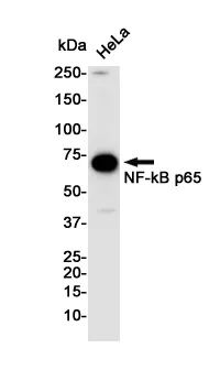
Western blot analysis of NF-KB p65 in Hela lysates using NF-KB p65 antibody.

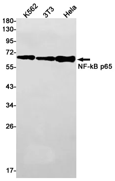
Western blot analysis of NF-KB p65 in K562, 3T3, Hela lysates using NF-KB p65 antibody.

Immunohistochemistry analysis of paraffin-embedded Human breast cancer using NF-KB p65 antibody. High-pressure and temperature Sodium Citrate pH 6.0 was used for antigen retrieval.

No documents available.