[KO Validated] Smad3 Rabbit mAb
![Western blot analysis of lysates from wild type and Smad3 knockout 293T cells, using [KO Validated] Smad3 Rabbit mAb at 1:1000 dilution.
Secondary antibody: HRP Goat Anti-Rabbit IgG at 1:10000 dilution.
Lysates/proteins: 25μg per lane.
Blocking buffer: 3% nonfat dry milk in TBST.
Detection: ECL Basic Kit .
Exposure time: 10s.](/storage/products/127167/ko-validated-smad3-rabbit-mab-ab23491-img-1.jpg)
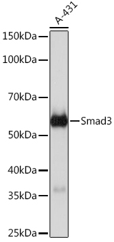
Western blot analysis of lysates from wild type and Smad3 knockout 293T cells, using [KO Validated] Smad3 Rabbit mAb at 1:1000 dilution. Secondary antibody: HRP Goat Anti-Rabbit IgG at 1:10000 dilution. Lysates/proteins: 25μg per lane. Blocking buffer: 3% nonfat dry milk in TBST. Detection: ECL Basic Kit . Exposure time: 10s.
| Catalog Number: | AB23491 |
| Conjugate: | Unconjugated |
| Size: | 100 ug |
| Concentration: | 1mg/ml |
| Host: | Rabbit |
| Isotype: | IgG |
| Clone: | ARC0490 |
| Immunogen: | Recombinant fusion protein containing a sequence corresponding to amino acids 119-219 of human Smad3 (NP_005893.1). |
| Reactivity: | Human,Mouse |
| Applications: | WB,1:500 - 1:1000 IHC-P,1:50 - 1:200 IF/ICC,1:50 - 1:200 IP,0.5μg-4μg antibody for 200μg-400μg extracts of whole cells |
| Molecular Weight: | 52kDa |
| Purification: | Affinity purification |
| Form: | liquid |
The SMAD family of proteins are a group of intracellular signal transducer proteins similar to the gene products of the Drosophila gene 'mothers against decapentaplegic' (Mad) and the C. elegans gene Sma. The SMAD3 protein functions in the transforming growth factor-beta signaling pathway, and transmits signals from the cell surface to the nucleus, regulating gene activity and cell proliferation. This protein forms a complex with other SMAD proteins and binds DNA, functioning both as a transcription factor and tumor suppressor. Mutations in this gene are associated with aneurysms-osteoarthritis syndrome and Loeys-Dietz Syndrome 3.
Applications
WB,1:500 - 1:1000 IHC-P,1:50 - 1:200 IF/ICC,1:50 - 1:200 IP,0.5μg-4μg antibody for 200μg-400μg extracts of whole cells
Immunogen
Recombinant fusion protein containing a sequence corresponding to amino acids 119-219 of human Smad3 (NP_005893.1).