KU70 Monoclonal Antibody

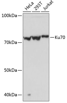
Western blot analysis of extracts of various cell lines, using Ku70 Rabbit mAb  at 1:1000 dilution.
Secondary antibody: HRP Goat Anti-Rabbit IgG (H+L) at 1:10000 dilution.
Lysates/proteins: 25ug per lane.
Blocking buffer: 3% nonfat dry milk in TBST.
Detection: ECL West Pico Plus
Exposure time: 1s.

Western blot analysis of extracts of various cell lines, using Ku70 Rabbit mAb at 1:1000 dilution. Secondary antibody: HRP Goat Anti-Rabbit IgG (H+L) at 1:10000 dilution. Lysates/proteins: 25ug per lane. Blocking buffer: 3% nonfat dry milk in TBST. Detection: ECL West Pico Plus Exposure time: 1s.

Catalog Number:MAB-94696
Conjugate:Unconjugated
Size:100 ug
Concentration:1mg/ml
Host:Rb
Isotype:IgG
Clone:EPR4027
Immunogen:Immunogen A synthetic peptide corresponding to a sequence within amino acids 500-609 of human Ku70.
Reactivity:Human, Mouse, Rat
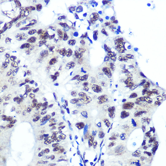
Applications:Western Blot: 1:500 - 1:2000 Immunohistochemistry: HC 1:50 - 1:200 Immunofluorescence: 1:50 - 1:200 Immunocytochemistry CC 1:50 - 1:200
Molecular Weight:70KDa
Purification:Aff. Pur.
Form:Liquid
Synonyms:CTC75; CTCBF; G22P1; KU70; ML8; TLAA

Applications

Western Blot: 1:500 - 1:2000 Immunohistochemistry: HC 1:50 - 1:200 Immunofluorescence: 1:50 - 1:200 Immunocytochemistry CC 1:50 - 1:200

Immunogen

Immunogen A synthetic peptide corresponding to a sequence within amino acids 500-609 of human Ku70.

Target Background

The p70/p80 autoantigen is a nuclear complex consisting of two subunits with molecular masses of approximately 70 and 80 kDa. The complex functions as a single-stranded DNA-dependent ATP-dependent helicase. The complex may be involved in the repair of nonhomologous DNA ends such as that required for double-strand break repair, transposition, and V(D)J recombination. High levels of autoantibodies to p70 and p80 have been found in some patients with systemic lupus erythematosus.

Synonyms:CTC75; CTCBF; G22P1; KU70; ML8; TLAA

Storage

Store at -20℃. Avoid freeze / thaw cycles.

Buffer

PBS with 0.01% thiomersal,0.05% BSA,50% glycerol,pH7.3.

Western blot analysis of extracts of various cell lines, using Ku70 Rabbit mAb at 1:1000 dilution. Secondary antibody: HRP Goat Anti-Rabbit IgG (H+L) at 1:10000 dilution. Lysates/proteins: 25ug per lane. Blocking buffer: 3% nonfat dry milk in TBST. Detection: ECL West Pico Plus Exposure time: 1s.

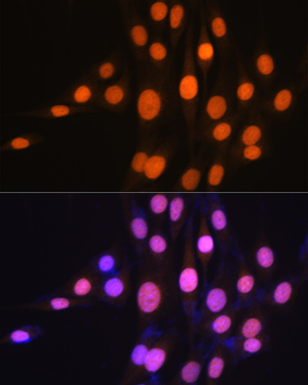
Immunofluorescence analysis of NIH-3T3 cells using Ku70 Rabbit mAb at dilution of 1:100 (40x lens). Blue: DAPI for nuclear staining.

Immunofluorescence analysis of NIH-3T3 cells using Ku70 Rabbit mAb at dilution of 1:100 (40x lens). Blue: DAPI for nuclear staining.

No documents available.