
Lamin A/C

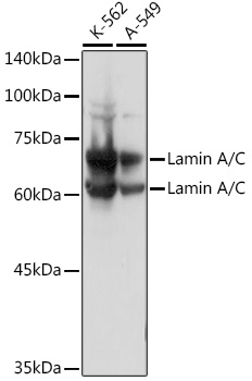
Western blot analysis of extracts of various cell lines, using Lamin A/C Rabbit pAb at 1:3000 dilution.
Secondary antibody: HRP Goat Anti-Rabbit IgG (H+L)  at 1:10000 dilution.
Lysates/proteins: 25ug per lane.
Blocking buffer: 3% nonfat dry milk in TBST.
Detection: ECL West Pico Plus.   
Exposure time: 1s.

Western blot analysis of extracts of various cell lines, using Lamin A/C Rabbit pAb at 1:3000 dilution. Secondary antibody: HRP Goat Anti-Rabbit IgG (H+L) at 1:10000 dilution. Lysates/proteins: 25ug per lane. Blocking buffer: 3% nonfat dry milk in TBST. Detection: ECL West Pico Plus. Exposure time: 1s.

Catalog Number:AB-90019
Conjugate:Unconjugated
Size:100 ug
Concentration:1mg/ml
Host:Rb
Isotype:IgG
Clone:POLY
Immunogen:Recombinant fusion protein containing a sequence corresponding to amino acids 403-572 of human Lamin C.
Reactivity:Human, Mouse, Rat
Applications:Western Blot: 1:500 - 1:2000 Immunohistochemistry: 1:50 - 1:200 Immunofluorescence: 1:50 - 1:200 ChIP: 1:20 - 1:100
Molecular Weight:63KDa
Purification:Aff. Pur.
Form:Liquid
Synonyms:LMNA; CDCD1; CDDC; CMD1A; CMT2B1; EMD2; FPL; FPLD; FPLD2; HGPS; IDC; LDP1; LFP; LGMD1B; ;LMN1;LMNC;LMNL1;MADA;PRO1;lamin;Lamin A/C

Applications

Western Blot: 1:500 - 1:2000 Immunohistochemistry: 1:50 - 1:200 Immunofluorescence: 1:50 - 1:200 ChIP: 1:20 - 1:100

Immunogen

Recombinant fusion protein containing a sequence corresponding to amino acids 403-572 of human Lamin C.

Target Background

The nuclear lamina consists of a two-dimensional matrix of proteins located next to the inner nuclear membrane. The lamin family of proteins make up the matrix and are highly conserved in evolution. During mitosis, the lamina matrix is reversibly disassembled as the lamin proteins are phosphorylated. Lamin proteins are thought to be involved in nuclear stability, chromatin structure and gene expression. Vertebrate lamins consist of two types, A and B. Alternative splicing results in multiple transcript variants. Mutations in this gene lead to several diseases: Emery-Dreifuss muscular dystrophy, familial partial lipodystrophy, limb girdle muscular dystrophy, dilated cardiomyopathy, Charcot-Marie-Tooth disease, and Hutchinson-Gilford progeria syndrome.

Synonyms:LMNA; CDCD1; CDDC; CMD1A; CMT2B1; EMD2; FPL; FPLD; FPLD2; HGPS; IDC; LDP1; LFP; LGMD1B; ;LMN1;LMNC;LMNL1;MADA;PRO1;lamin;Lamin A/C

Storage

Store at -20℃. Avoid freeze / thaw cycles.

Buffer

PBS with 0.02% sodium azide,50% glycerol,pH7.3

Western blot analysis of extracts of various cell lines, using Lamin A/C Rabbit pAb at 1:3000 dilution. Secondary antibody: HRP Goat Anti-Rabbit IgG (H+L) at 1:10000 dilution. Lysates/proteins: 25ug per lane. Blocking buffer: 3% nonfat dry milk in TBST. Detection: ECL West Pico Plus. Exposure time: 1s.

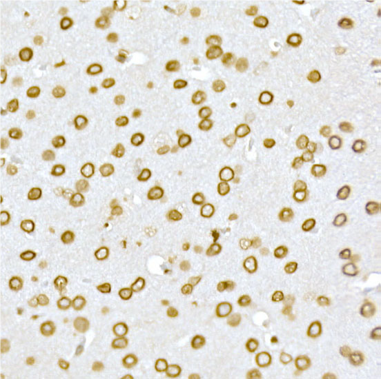
Immunohistochemistry of paraffin embedded mouse brain using [KO Validated] Lamin A/C Rabbit pAb at dilution of 1:50 (40x ns). Perform high pressure antigen retrieval with 10 mM citrate buffer pH 6.0 before commencing with IHC staining protocol.

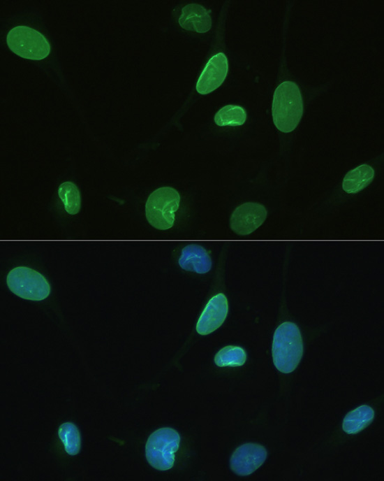
Immunofluorescence analysis of U2OS using Lamin A/C antibody at dilution of 1:100. Blue: DAPI for nuclear staining.

No documents available.