
Lamin A/C

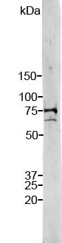
Strip blot of crude HeLa cell extract stained with RPCA-LaminAC. Note two strong and clean bands at 74 kDa and 65 kDa, corresponding to Lamin A and C.

Strip blot of crude HeLa cell extract stained with RPCA-LaminAC. Note two strong and clean bands at 74 kDa and 65 kDa, corresponding to Lamin A and C.

Catalog Number:MAB-94259
Conjugate:Unconjugated
Size:100 ug
Concentration:1mg/ml
Host:Ms
Isotype:IgG1
Clone:4C4
Immunogen:Full length human Lamin A purified form E. coli.
Reactivity:Mammalian
Applications:Western Blot: 1:10000 Immunohistochemistry: 1:1000 Immunocytochemistry 1:1000 Immunofluorescence: 1000
Molecular Weight:65, 74 kDa
Purification:Purified
Form:Liquid

Applications

Western Blot: 1:10000 Immunohistochemistry: 1:1000 Immunocytochemistry 1:1000 Immunofluorescence: 1000

Immunogen

Full length human Lamin A purified form E. coli.

Target Background

The Lamin proteins are members of the intermediate filament protein family but are located inside the nucleus rather than in the cytoplasm (1). The lamins function as skeletal components tightly associated with the inner nuclear membrane. Originally the proteins of the nuclear cytoskeleton were named Lamin A, B and C, from top to bottom as visualized on SDS-PAGE gels. Subsequently, it was found that Lamins A and C were coded for by a single gene (2), while the Lamin B band may contain two proteins encoded by two genes now called Lamin B1 and Lamin B2. Lamin A has a mass of about 74 kDa while Lamin C is 65 kDa. The Lamin A protein includes a C-terminal segment of 98 amino acids missing from Lamin C, while Lamin C has a unique C-terminal 6 amino acid peptide not present in Lamin A. Apart from these regions Lamin A and C are identical so that antibodies raised against either protein are likely to cross react with the other. Lamin polymerization and depolymerization is regulated by phosphorylation by cyclin dependent protein kinase 1 (CDK1), the key component of “maturation promoting factor“, the central regulator of cell division. Activity of this kinase increases during cell division and is responsible for the breakdown of the nuclear lamina.

Storage

Store at 4°C. For long term storage, store at -20°C. Avoid freeze / thaw cycles.

Buffer

10mM sodium azide preservative

Strip blot of crude HeLa cell extract stained with RPCA-LaminAC. Note two strong and clean bands at 74 kDa and 65 kDa, corresponding to Lamin A and C.

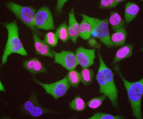
HeLa cells staing with RPCA-LaminAC (red) counterstained with monoclonal antibody to HSP27, (green) and DNA (blue). The RPCA-LaminAC antibody reveals strong nuclear lamina staining, while the MCA-6H11 antibody reveals strong cytoplasmic staining. Since both DNA (blue) and Lamin A/C (red) are associated with the nuclear compartment, this region appears crimson in this image.

No documents available.