
Lamin A/C

Western blot analysis of cytosolic or nuclear enriched fractions of
cell lines probed with chicken pAb to lamin A/C, ,
dilution 1:1,000 in green: [1] protein standard (red), [2] HeLa
cytosol, HeLa nuclear, [4] NIH-3T3 cytosol, and [5] NIH-3T3
nuclear fractions. Two strong bands at 65kDa and 74kDa correspond to lamin A and lamin C proteins respectively, detected exclusively in the nuclear fractions. The same blot was simultaneously probed with
mouse mAb to GAPDH, in red. The single band at 37kDa
represents GAPDH protein which is expressed predominantly in the
cytosolic fractions.

Western blot analysis of cytosolic or nuclear enriched fractions of cell lines probed with chicken pAb to lamin A/C, , dilution 1:1,000 in green: [1] protein standard (red), [2] HeLa cytosol, HeLa nuclear, [4] NIH-3T3 cytosol, and [5] NIH-3T3 nuclear fractions. Two strong bands at 65kDa and 74kDa correspond to lamin A and lamin C proteins respectively, detected exclusively in the nuclear fractions. The same blot was simultaneously probed with mouse mAb to GAPDH, in red. The single band at 37kDa represents GAPDH protein which is expressed predominantly in the cytosolic fractions.

Catalog Number:AB- 83935
Conjugate:Unconjugated
Size:100 ug
Concentration:1mg/ml
Host:Ch
Isotype:IgY
Clone:POLY
Immunogen:Full length recombinant human lamin A protein expressed in and purified from E. coli.
Reactivity:Hu, Rt, Ms,Ho dg mk
Applications:Western Blot: 1:2,000 Immunofluorescence: 1:1,000 Immunocytochemistry: 1:1,000
Molecular Weight:65kDa, 74kDa
Purification:Purified
Form:Liquid

Applications

Western Blot: 1:2,000 Immunofluorescence: 1:1,000 Immunocytochemistry: 1:1,000

Immunogen

Full length recombinant human lamin A protein expressed in and purified from E. coli.
Target Background

Lamin A and lamin C are members of the intermediate filament protein family and are located in the nucleus where they function as skeletal components of the inner nuclear membrane (1). The two proteins are generated by alternate transcription from the single LMNA gene. Lamin A has a molecular weight of about 74kDa while lamin C is 65kDa. The lamin A protein includes a C-terminal segment of 98 amino acids missing from lamin C, while lamin C has a unique C-terminal 6 amino acid peptide not present in lamin A. As a result antibodies raised against lamin A are almost certain bind to lamin C. During cell division the nuclear lamina breaks down and lamin A/C containing filaments depolymerize, this being regulated by phosphorylation by cyclin dependent protein kinase 1. Mutations in the lamin A/C gene are associated with several serious human diseases, including Emery-Dreifuss muscular dystrophy, familial partial lipodystrophy, limb girdle muscular dystrophy, dilated cardiomyopathy, Charcot-Marie-Tooth disease type 2B1, Hutchinson-Gilford progeria syndrome and Hutchinson-Gilford progeria syndrome.

Storage

Stable at 4°C for one year.

Buffer

Supplied as an aliquot of concentrated IgY preparation plus 5mM NaN3

Western blot analysis of cytosolic or nuclear enriched fractions of cell lines probed with chicken pAb to lamin A/C, , dilution 1:1,000 in green: [1] protein standard (red), [2] HeLa cytosol, HeLa nuclear, [4] NIH-3T3 cytosol, and [5] NIH-3T3 nuclear fractions. Two strong bands at 65kDa and 74kDa correspond to lamin A and lamin C proteins respectively, detected exclusively in the nuclear fractions. The same blot was simultaneously probed with mouse mAb to GAPDH, in red. The single band at 37kDa represents GAPDH protein which is expressed predominantly in the cytosolic fractions.

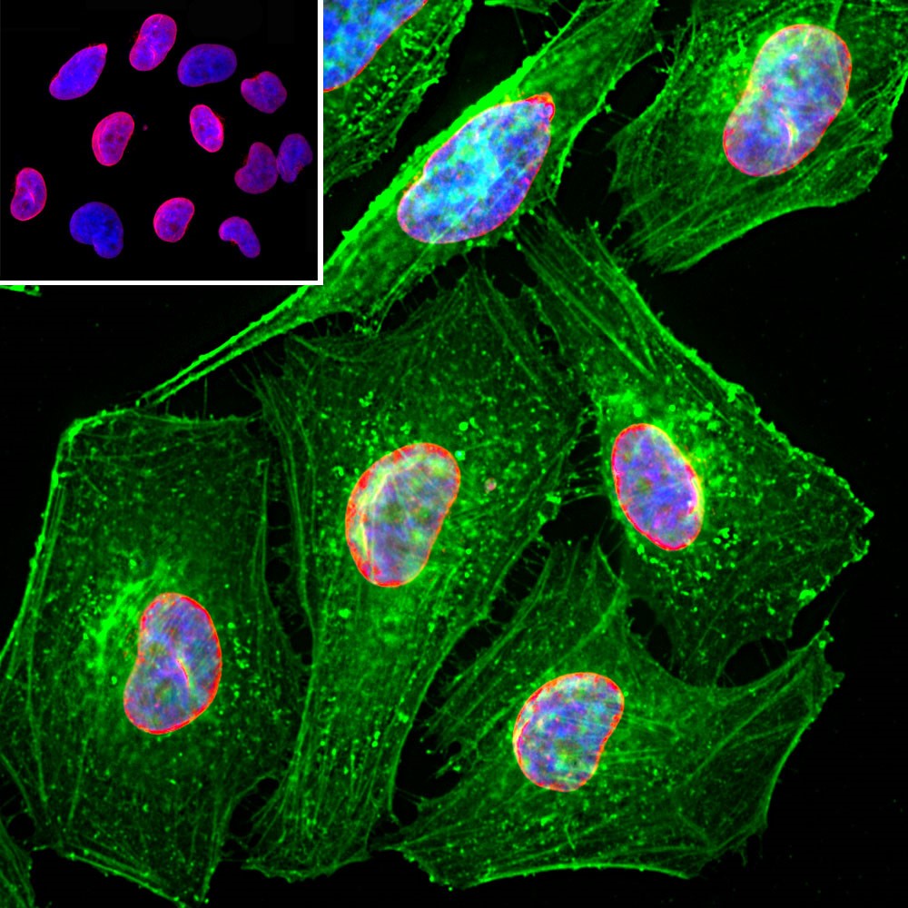
Immunofluorescent analysis of HeLa cells stained with chicken pAb to lamin A/C, dilution 1:2,000 in red, and costained with mouse mAb to actin, dilution 1:500, in green. The blue is Hoechst staining of nuclear DNA. The LaminAC antibody specifically labels the nuclear lamina, while the actin antibody stains the submembranous actin-rich cytoskeleton, stress fibers and bundles of actin associated with cell adhesion sites.

No documents available.