
Laminin Rabbit Polyclonal Antibody

Western blot analysis of different tissue lysates using rabbit pAb to laminin, dilution 1:5,000 in green: [1] protein standard (red), [2] rat brain, [3] rat spinal cord, and [4] cow spinal cord lysate. The strong band above the 280kDa mark corresponds to full length laminin proteins. Smaller proteolytic fragments of laminin are also detected with this antibody.

Western blot analysis of different tissue lysates using rabbit pAb to laminin, dilution 1:5,000 in green: [1] protein standard (red), [2] rat brain, [3] rat spinal cord, and [4] cow spinal cord lysate. The strong band above the 280kDa mark corresponds to full length laminin proteins. Smaller proteolytic fragments of laminin are also detected with this antibody.

Catalog Number:AB- 83936
Conjugate:Unconjugated
Size:100 ug
Concentration:1mg/ml
Host:Rabbit
Isotype:IgG
Clone:POLY
Immunogen:Laminin 111 from mouse EHS sarcoma cells obtained from Sigma-Aldrich.
Reactivity:Human, Rat, Mouse
Applications:Western Blot: 1:5,000 Immunofluorescence: 1:1,000-5,000 Immunocytochemistry: 1:1,000-5,000 Immunohistochemistry: 1:1,000-5,000
Molecular Weight:440kDa, 220kDa, 158kDa
Purification:Aff. Pur.
Form:Liquid

Applications

Western Blot: 1:5,000 Immunofluorescence: 1:1,000-5,000 Immunocytochemistry: 1:1,000-5,000 Immunohistochemistry: 1:1,000-5,000

Immunogen

Laminin 111 from mouse EHS sarcoma cells obtained from Sigma-Aldrich.
Target Background

Laminins are high-molecular weight proteins and major components of the extracellular matrix. They are an important part of the basal lamina a protein network typically separating cells and tissues of different embryonic origin. Laminin was first isolated biochemically from Engelbreth-Holm-Swarm (EHS) mouse sarcoma cells, a cell line which produces large amounts of extracellular matrix material (1,2). Laminins are heterotrimeric proteins that contain one of five α-chains, one of three β-chains and one of three γ-chains (3-4). The EHS sarcoma cell derived laminin was originally named laminin 1, composed of α1β1γ1 polypeptides (5), though a later nomenclature named this form laminin 111, the numbers indicating the content of α, β and γ gene products (6). The distribution of the different laminin isoforms is developmental time and tissue-specific and laminin-111 expressed in the embryonic epithelium, but is also expressed in adult kidney, liver, testis, ovaries and brain blood vessels (7). Genetic ablation of laminin-111 results in embryonic death (8), and point mutations in the human α1 gene resulted in Poretti Boltshauser Syndrome, a rare disorder associated with cerebellar abnormalities, ataxia, cognitive problems and other issues. Injection of laminin–111 into muscle appeared to be a promising therapy in a mouse model Duchenne muscular dystrophy (DMD, 9). However transgenic overexpression of the laminin α1 protein appeared to have no beneficial affect also in a mouse DMD model (10). The Laminin antibody was made against laminin 111 isolated biochemically from mouse Engelbreth-Holm-Swarm (EHS) sarcoma cells (1,2). The antibody recognizes the several different laminin polypeptides on western blots and is a very useful reagent for visualizing basal lamina in mature and developing tissues. It is particularly useful in the CNS, allowing visualization of the basal lamina of endothelia, an important part of the blood brain barrier. Mouse select image above left for larger view.

Storage

Storage for short term at 4°C recommended, for longer term at -20°C.

Buffer

Affinity purified antibody at 1mg/mL in 50% PBS, 50% glycerol plus 5mM NaN3

Western blot analysis of different tissue lysates using rabbit pAb to laminin, dilution 1:5,000 in green: [1] protein standard (red), [2] rat brain, [3] rat spinal cord, and [4] cow spinal cord lysate. The strong band above the 280kDa mark corresponds to full length laminin proteins. Smaller proteolytic fragments of laminin are also detected with this antibody.

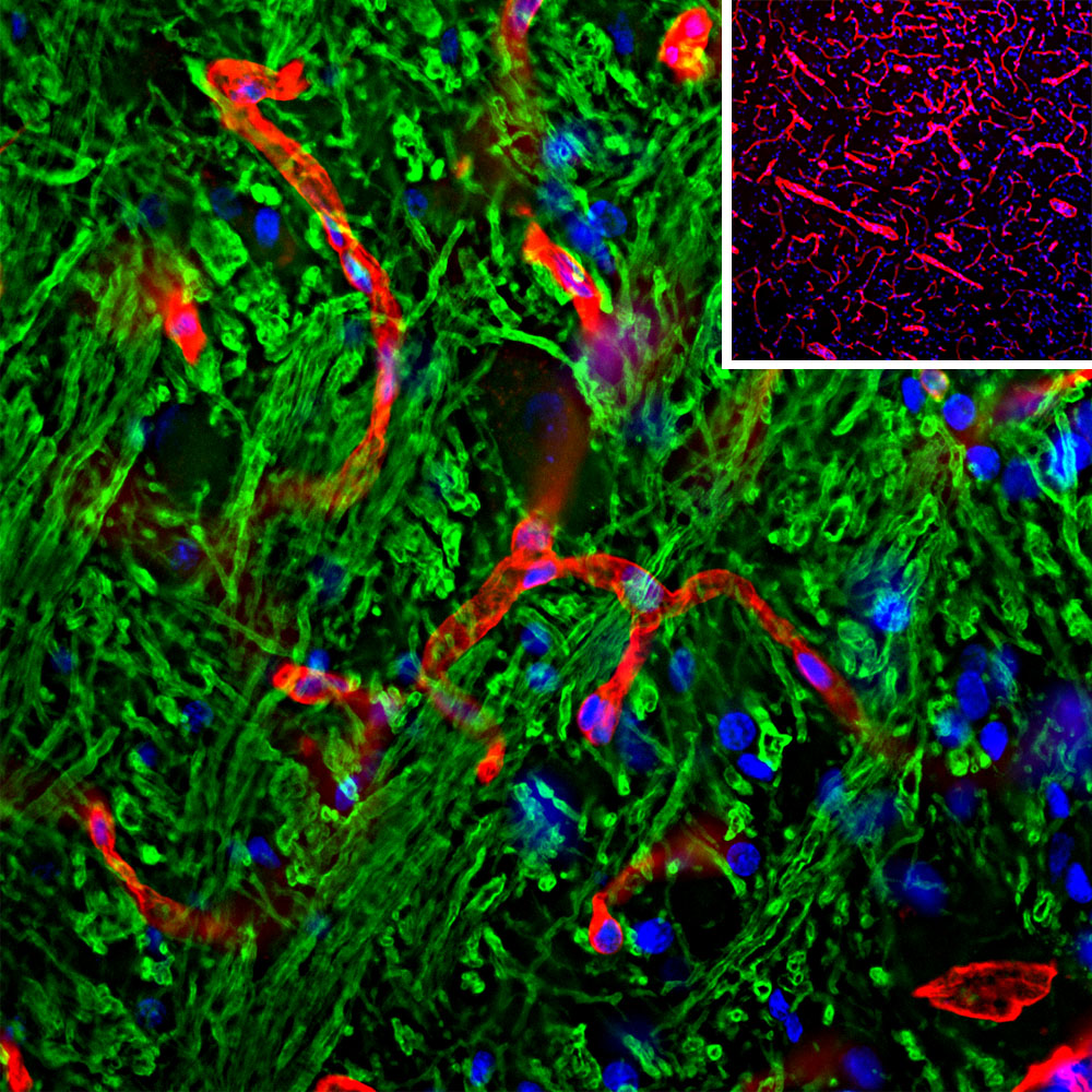
Immunohistological analysis of brain stem section stained with rabbit pAb to laminin, dilution 1:1,000 in red, and costained with chicken pAb to myelin basic protein (MBP), dilution 1:5,000 in green. The blue is DAPI staining of nuclear DNA. Following transcardial perfusion of rat with 4% paraformaldehyde, brain was post fixed for 24 hours, cut to 45μM, and free-floating sections were stained with the above antibodies. The laminin antibody is an excellent marker of basement membranes surrounding blood vessels, while the MBP antibody stains the myelin sheathes around axons.

No documents available.