LAMP1 / CD107a Mouse Monoclonal Antibody
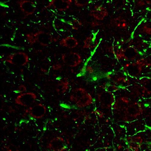
Immunohistochemistry staining of CD107a (red) in tissue sections of murine brain expressing GFP in some of its neurons (green). Mouse monoclonal antibody H4A3 (anti-CD107a) was detected with goat anti-mouse IgG1 conjugated with Alexa Fluor 555.

Immunohistochemistry staining of CD107a (red) in tissue sections of murine brain expressing GFP in some of its neurons (green). Mouse monoclonal antibody H4A3 (anti-CD107a) was detected with goat anti-mouse IgG1 conjugated with Alexa Fluor 555.

Catalog Number:MAB-94450
Conjugate:Unconjugated
Size:100 ug
Concentration:1mg/ml
Host:Mouse
Isotype:IgG1 kappa
Clone:H4A3
Immunogen:Human PBMC
Reactivity:Human, Mouse
Applications:Western blotting: Recommended dilution: 1 μg/ml. Immunohistochemistry: Recommended dilution: 2-8 μg/ml. Flow cytometry: Intracellular and extracellular staining; recommended dilution: 1-12 µg/ml.
Molecular Weight:100-120kDa
Purification:Purified
Form:Liquid
Synonyms:LAMP-1, LAMPA

Applications

Western blotting: Recommended dilution: 1 μg/ml. Immunohistochemistry: Recommended dilution: 2-8 μg/ml. Flow cytometry: Intracellular and extracellular staining; recommended dilution: 1-12 µg/ml.

Immunogen

Human PBMC
Target Background

CD107a (lysosome-associated membrane protein-1, LAMP-1), together with LAMP-2, is a major constituent of lysosomal membrane, 1-2% of total CD107a is found also on the plasma membrane. The LAMP proteins are involved in lysosome biogenesis and are required for fusion of lysosomes with phagosomes. Increased CD107a immunoreactivity is observed in neurones, and in glial cells surrounding senile plaques in Alzheimers disease cases and is localized mainly in medullary epithelial cells, single macrophages and lymphocytes in acute thymic involution. CD107a is a good marker of mast cell activation.

Synonyms:LAMP-1, LAMPA

Storage

Store at 2-8°C. Do not freeze.

Buffer

Phosphate buffered saline (PBS), pH 7.4

Immunohistochemistry staining of CD107a (red) in tissue sections of murine brain expressing GFP in some of its neurons (green). Mouse monoclonal antibody H4A3 (anti-CD107a) was detected with goat anti-mouse IgG1 conjugated with Alexa Fluor 555.

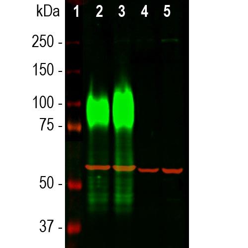
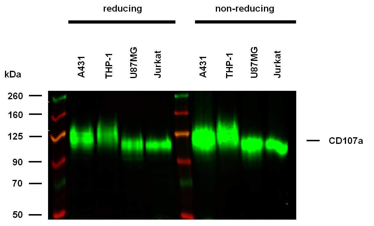
<b>Anti-Hu CD107a Purified (clone H4A3) works in WB application under reducing and non-reducing conditions.</b> Western blotting analysis was performed on whole cell extracts (RIPA lysis buffer) of A431, THP-1, U87MG, and Jurkat cell lines, mixed and heated (100°C, 5 min) with reducing and non-reducing SDS-loading buffer. Samples were resolved using 7% SDS-PAGE gel. Nitrocellulose membrane blot was probed with mouse IgG1 monoclonal antibody H4A3 (1 µg/ml), followed by IRDye 800CW Goat-anti-Mouse IgG (green). Multiplex fluorescent Western blot detection was performed. CD107a molecules were detected at 120 kDa in all tested cell lines under both reducing and non-reducing conditions.

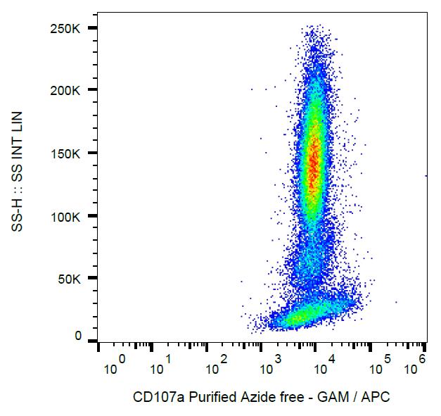
Flow cytometry analysis (intracellular staining) of human peripheral blood cells with anti-CD107a (H4A3) azide free, GAM-APC.

Western blot analysis of different cell lysates using mouse mAb to LAMP1, dilution 1:10,000 in green. Cells were maintained under normal conditions (Ct), or treated with 50

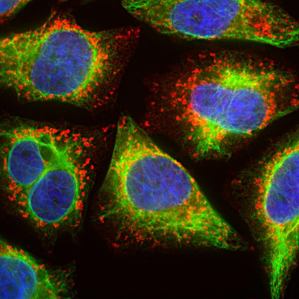
Immunofluorescent analysis of HeLa cells stained with mouse mAb to LAMP1, dilution 1:500 in red, and co-stained with chicken pAb to vimentin, dilution 1:10,000, in green. The blue is DAPI staining of nuclear DNA. The cells were treated with 50

No documents available.