
LRRK2

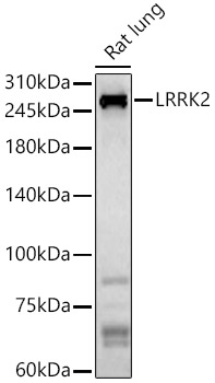
Western blot analysis of Rat lung, using LRRK2 antibody at 1:800 dilution.
Secondary Antibody: HRP Goat Anti-Rabbit IgG (H+L) at 1:10000 dilution.
Lysates/proteins: 25ug per lane.
Blocking buffer: 3% nonfat dry milk in TBST.
Detection: ECL Basic Kit.
Exposure time: 180s.

Western blot analysis of Rat lung, using LRRK2 antibody at 1:800 dilution. Secondary Antibody: HRP Goat Anti-Rabbit IgG (H+L) at 1:10000 dilution. Lysates/proteins: 25ug per lane. Blocking buffer: 3% nonfat dry milk in TBST. Detection: ECL Basic Kit. Exposure time: 180s.

Catalog Number:AB-82808
Size:100 ug
Concentration:1mg/ml
Host:Rabbit
Isotype:IgG
Clone:POLY
Immunogen:Recombinant fusion protein containing a sequence corresponding to amino acids 750-910 of human LRRK2
Reactivity:Hu, Ms, Rt
Applications:WB 1:500 - 1:1000 IHC 1:50 – 1:200 IF 1:50 – 1:200 ICC 1:50 – 1:200
Molecular Weight:290 kDa
Purification:Aff. Pur.
Form:Liquid
Synonyms:PARK8; RIPK7; ROCO2; AURA17; DARDARIN

Applications

WB 1:500 - 1:1000 IHC 1:50 – 1:200 IF 1:50 – 1:200 ICC 1:50 – 1:200

Immunogen

Recombinant fusion protein containing a sequence corresponding to amino acids 750-910 of human LRRK2

Target Background

This gene is a member of the leucine-rich repeat kinase family and encodes a protein with an ankryin repeat region, a leucine-rich repeat (LRR) domain, a kinase domain, a DFG-like motif, a RAS domain, a GTPase domain, a MLK-like domain, and a WD40 domain. The protein is present largely in the cytoplasm but also associates with the mitochondrial outer membrane. Mutations in this gene have been associated with Parkinson disease-8.

Synonyms:PARK8; RIPK7; ROCO2; AURA17; DARDARIN

Storage

Store at -20℃. Avoid freeze / thaw cycles

Buffer

PBS with 0.01% thiomersal,50% glycerol, pH7.3.

Western blot analysis of Rat lung, using LRRK2 antibody at 1:800 dilution. Secondary Antibody: HRP Goat Anti-Rabbit IgG (H+L) at 1:10000 dilution. Lysates/proteins: 25ug per lane. Blocking buffer: 3% nonfat dry milk in TBST. Detection: ECL Basic Kit. Exposure time: 180s.

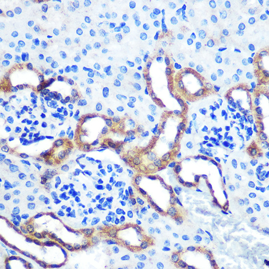
Immunohistochemistry of paraffinembedded Mouse kidney using LRRK2 Rabbit pAb at dilution of 1:100 (40x lens). Perform microwave antigen retrieval with 10 mM PBS buffer pH 7.2 before commencing with IHC staining protocol

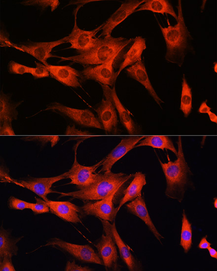
Immunofluorescence analysis of C2C12 cells using LRRK2 Rabbit pAb at dilution of 1:100 (40x lens). Blue: DAPI for nuclear staining.

No documents available.