
MAP-2ab

Western blot analysis of different tissue lysates using rabbit pAb to
microtubule associated protein 2A and 2B (MAP2A/B), MAP2A/
B, dilution 1:50,000 in green: [1] protein standard (red), [2]
rat brain, [3] mouse brain, and [4] caw cortex. Strong band at about
280kDa mark corresponds to the two major high molecular weight
MAP2 isoforms referred to as MAP2A and MAP2B. Smaller fragments
of these isoforms are also detected on the blot, but the MAP2C and
MAP2D isoforms are not.

Western blot analysis of different tissue lysates using rabbit pAb to microtubule associated protein 2A and 2B (MAP2A/B), MAP2A/ B, dilution 1:50,000 in green: [1] protein standard (red), [2] rat brain, [3] mouse brain, and [4] caw cortex. Strong band at about 280kDa mark corresponds to the two major high molecular weight MAP2 isoforms referred to as MAP2A and MAP2B. Smaller fragments of these isoforms are also detected on the blot, but the MAP2C and MAP2D isoforms are not.

Catalog Number:AB-84327
Conjugate:Unconjugated
Size:100 ug
Concentration:1mg/ml
Host:Rb
Isotype:IgG
Clone:POLY
Immunogen:Recombinant human projection domain sequences, amino acids 377-1505.
Reactivity:Hu, Ms, Rt
Applications:Western Blot: 1:1,000 Immunofluorescence: 1:500-1:1000 Immunocytochemistry: 1;500-1:1000 Immunohistochemistry: 1:500-1:1000
Molecular Weight:~280kDa
Purification:Aff. Pur.
Form:Liquid

Applications

Western Blot: 1:1,000 Immunofluorescence: 1:500-1:1000 Immunocytochemistry: 1;500-1:1000 Immunohistochemistry: 1:500-1:1000

Immunogen

Recombinant human projection domain sequences, amino acids 377-1505.
Target Background

Microtubules are 25nm diameter protein rods found in most kinds of eukaryotic cells and are associated with a family of proteins called microtubule associated proteins (MAPs). MAPs play a crucial role in the regulation of microtubule dynamics and interactions in vivo. MAP2 was originally named as one of the higher molecular weight MAPs with an SDS-PAGE molecular weight of about 280kDa (1-3). There is a single mammalian MAP2 gene which may generates two high molecular weight proteins of ~280kDa name MAP2A and MAP2B and multiple lower molecular weight forms usually named MAP2C and MAP2D which run on SDS-PAGE gels at 60-70kDa. The lower molecular weight forms are found in neurons early in development, but later are replaced by the higher molecular weight forms (2). The MAP2A and MAP2B forms include a protein insert missing in MAP2C and MAP2D which forms fine filamentous protrusions from the sides of brain microtubules referred to as the "projection domain". This antibody was made against the projection domain sequences and so is specific for MAP2A and MAPB. MAP2 isoforms are expressed only in neuronal perikarya and dendrites so appropriate antibodies are used to identify neurons and dendrites in cell culture and sections. The MAP2 antibody was made against a mixture of recombinant human projection domain sequences, amino acids 377-1505. It binds to the MAP2A and MAP2B isoforms but not the MAP2C and MA2PD which lack projection domain sequences

Storage

Stable at 4°C for one year, for longer term store at -20°C

Buffer

Purified antibody at 1mg/mL in 50% PBS, 50% glycerol plus 5mM NaN3

Western blot analysis of different tissue lysates using rabbit pAb to microtubule associated protein 2A and 2B (MAP2A/B), MAP2A/ B, dilution 1:50,000 in green: [1] protein standard (red), [2] rat brain, [3] mouse brain, and [4] caw cortex. Strong band at about 280kDa mark corresponds to the two major high molecular weight MAP2 isoforms referred to as MAP2A and MAP2B. Smaller fragments of these isoforms are also detected on the blot, but the MAP2C and MAP2D isoforms are not.

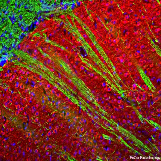
Immunofluorescent analysis of rat brain striatum stained with rabbit pAb to microtubule associated protein 2A and 2B isotypes (MAP2A/B), MAP2A/B, dilution 1:10,000 in red, and costained with chicken pAb to myelin basic protein (MBP), dilution 1:5,000 in green. The blue is Hoechst staining of nuclear DNA. The MAP2A/B antibody stains dendrites and perikarya of neurons, while the MBP antibody labels oligodendrocytes and myelin sheathes around axons.

No documents available.