MAP1LC3A / MAP1LC3B Rabbit Polyclonal Antibody
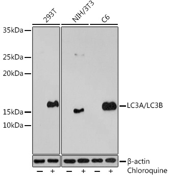
Western blot analysis of extracts of various cell lines, using LC3A/LC3B antibody at 1:500
dilution.293T cells were treated by Chloroquine (50 μM) at 37℃ for 20 hours.NIH/3T3 cells were
treated by Chloroquine (50 μM) at 37℃ for 20 hours.C6 cells were treated by Chloroquine (50 μM) at
37℃ for 20 hours.
Secondary antibody: HRP Goat Anti-Rabbit IgG (H+L) at 1:10000 dilution.
Lysates/proteins: 25μg per lane.
Blocking buffer: 3% BSA.
Detection: ECL West Pico Plus.
Exposure time: 3min.

Western blot analysis of extracts of various cell lines, using LC3A/LC3B antibody at 1:500 dilution.293T cells were treated by Chloroquine (50 μM) at 37℃ for 20 hours.NIH/3T3 cells were treated by Chloroquine (50 μM) at 37℃ for 20 hours.C6 cells were treated by Chloroquine (50 μM) at 37℃ for 20 hours. Secondary antibody: HRP Goat Anti-Rabbit IgG (H+L) at 1:10000 dilution. Lysates/proteins: 25μg per lane. Blocking buffer: 3% BSA. Detection: ECL West Pico Plus. Exposure time: 3min.

Catalog Number:AB-83557
Conjugate:Unconjugated
Size:100 ul
Concentration:1mg/ml
Host:Rabbit
Isotype:IgG
Clone:POLY
Immunogen:A synthetic peptide corresponding to a sequence within amino acids 1-60 of human LC3A
Reactivity:Human,Mouse, Rat
Applications:Western Blot: 1:500 - 1:2000Immunohistochemistry(paraffin-embedded tissues): 1:50 - 1:200Immunofluorescence:Immunocytochemistry: 1:50 - 1:200
Molecular Weight:14-16kDa
Purification:Purified
Form:Liquid
Synonyms:MAP1LC3B;ATG8F;LC3B;MAP1A/1BLC3;MAP1LC3B-a;MAP1LC3A;ATG8E;LC3;LC3A;MAP1ALC3;MAP1BLC3;LC3A/LC3B

Applications

Western Blot: 1:500 - 1:2000Immunohistochemistry(paraffin-embedded tissues): 1:50 - 1:200Immunofluorescence:Immunocytochemistry: 1:50 - 1:200

Immunogen

A synthetic peptide corresponding to a sequence within amino acids 1-60 of human LC3A
Target Background

MAP1A and MAP1B are microtubule-associated proteins which mediate the physical interactions between microtubules and components of the cytoskeleton. MAP1A and MAP1B each consist of a heavy chain subunit and multiple light chain subunits. The protein encoded by this gene is one of the light chain subunits and can associate with either MAP1A or MAP1B. Two transcript variants encoding different isoforms have been found for this gene. The expression of variant 1 is suppressed in many tumor cell lines, suggesting that may be involved in carcinogenesis.

Synonyms:MAP1LC3B;ATG8F;LC3B;MAP1A/1BLC3;MAP1LC3B-a;MAP1LC3A;ATG8E;LC3;LC3A;MAP1ALC3;MAP1BLC3;LC3A/LC3B

Storage

Store at -20℃. Avoid freeze / thaw cycles.

Buffer

PBS with 0.01% thimerosal,50% glycerol,pH7.3.

Western blot analysis of extracts of various cell lines, using LC3A/LC3B antibody at 1:500 dilution.293T cells were treated by Chloroquine (50 μM) at 37℃ for 20 hours.NIH/3T3 cells were treated by Chloroquine (50 μM) at 37℃ for 20 hours.C6 cells were treated by Chloroquine (50 μM) at 37℃ for 20 hours. Secondary antibody: HRP Goat Anti-Rabbit IgG (H+L) at 1:10000 dilution. Lysates/proteins: 25μg per lane. Blocking buffer: 3% BSA. Detection: ECL West Pico Plus. Exposure time: 3min.

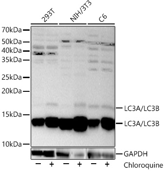
Western blot analysis of various lysates, using LC3A/LC3B Rabbit pAb at 1:2000 dilution.293T,NIH/3T3 and C6 cells were treated by Chloroquine (50 μM) at 37℃ for 20 hours. Secondary antibody: HRP Goat Anti-Rabbit IgG (H+L) at 1:10000 dilution. Lysates/proteins: 25ug per lane. Blocking buffer: 3% nonfat dry milk in TBST. Detection: ECL West Pico Plus. Exposure time: 30s.

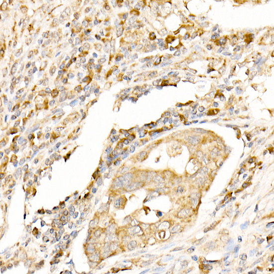
Immunohistochemistry analysis of paraffin-embedded human colon carcinoma using LC3A/LC3B Rabbit pAb at dilution of 1:100 (40x lens).Perform high pressure antigen retrieval with 10 mM citrate buffer pH 6.0 before commencing with IHC staining protocol.

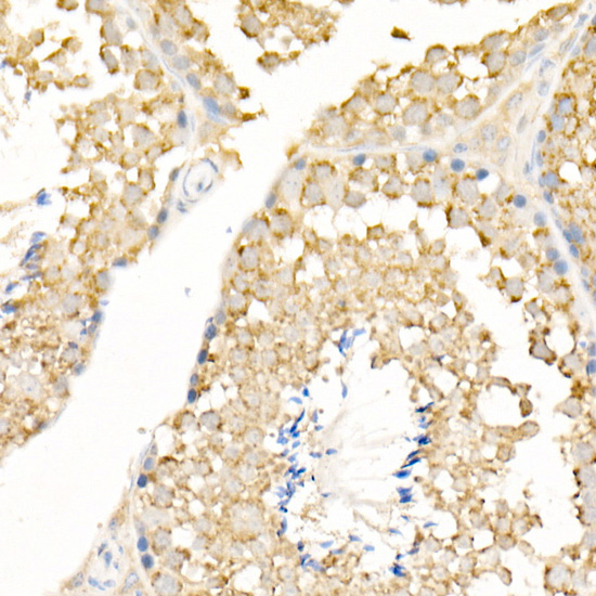
Immunohistochemistry analysis of paraffin-embedded mouse testis using LC3A/LC3B Rabbit pAb at dilution of 1:100 (40x lens).Perform high pressure antigen retrieval with 10 mM citrate buffer pH 6.0 before commencing with IHC staining protocol.

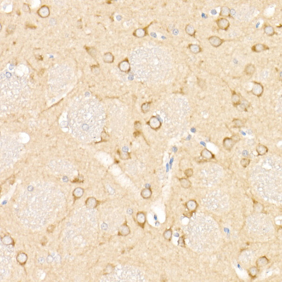
Immunohistochemistry analysis of paraffin-embedded rat brain using LC3A/LC3B Rabbit pAb at dilution of 1:100 (40x lens).Perform high pressure antigen retrieval with 10 mM citrate buffer pH 6.0 before commencing with IHC staining protocol.

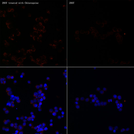
Immunofluorescence analysis of 293T Treated with Chloroquine and 293T cells using LC3A/LC3B Rabbit pAb at dilution of 1:100 (40x lens). Blue: DAPI for nuclear staining.

No documents available.