
mCherry Mouse Monoclonal Antibody

Western blot analysis of HEK293 cell lysates, and recombinant protein solutions using mouse mAb to mCherry, dilution
1:1,000, in green [1] protein standard, [2] HEK293, [3] HEK293 cells transfected with mCherry-HA construct, [4] mCherry recombinant protein, [5] GFP recombinant protein, and [6] HEK293 transfected with GFP construct. Major band at about 30kDa corresponds to mCherry protein. mCherry antibody does not react with GFP protein. The same blot was simultaneously probed with chicken pAb to HSP60, dilution 1:5,000 in red which reveals band at 60kDa seen only in cell lysates.

Western blot analysis of HEK293 cell lysates, and recombinant protein solutions using mouse mAb to mCherry, dilution 1:1,000, in green [1] protein standard, [2] HEK293, [3] HEK293 cells transfected with mCherry-HA construct, [4] mCherry recombinant protein, [5] GFP recombinant protein, and [6] HEK293 transfected with GFP construct. Major band at about 30kDa corresponds to mCherry protein. mCherry antibody does not react with GFP protein. The same blot was simultaneously probed with chicken pAb to HSP60, dilution 1:5,000 in red which reveals band at 60kDa seen only in cell lysates.

Catalog Number:MAB-94019
Conjugate:Unconjugated
Size:100 ug
Concentration:1mg/ml
Host:Mouse
Isotype:IgG2a heavy, κ light
Clone:1C51
Immunogen:Full length recombinant protein
Reactivity:All Species
Applications:Western Blot: 1:2,000 Immunofluorescence: 1:500 Immunohisotchemistry: 1:500
Molecular Weight:~28kDa
Purification:Purified
Form:Liquid

Applications

Western Blot: 1:2,000 Immunofluorescence: 1:500 Immunohisotchemistry: 1:500

Immunogen

Full length recombinant protein
Target Background

mCherry protein is derived from a natural product, DsRed, originally isolated as a red fluorescent protein from the coral of the genus Discosoma (1). As with other natural fluorescent proteins of Cnidarians (jelly fish, sea anemones, and corals), the natural form of the protein forms stable tetramers in vivo. DsRed was engineered to improve its spectral properties and also prevent ultimerization in the Tsien lab, where much work on fluorescent proteins was performed (2). Roger Tsien, along with Martin Chalfie, and Osamyu Shinomura shared the 2008 Nobel prize in chemistry for the discovery and exploitation of Cnidarian fluorescent proteins. Several further cycles of mutation, directed modification and evolutionary selection produced mCherry, which is monomeric and has an excitation maximum at 587nm and emission maximum at 610nm (3). The protein is widely used as a fluorescent tracer in transfection, transgenic, photobleaching and FRET type experiments. The prototype for these fluorescent proteins is Green Fluorescent Protein (GFP), which is a ~27kDa protein isolated originally from the jellyfish Aequoria victoria (4). The mCherry protein is similar in size and general structural properties to GFP (5,6), but, obviously, produces a red rather than a green fluorochrome. As with GFP, mCherry becomes fluorescent due to intrinsic properties requiring only molecular oxygen and so can be readily expressed in a variety of systems.

Storage

Stable at 4°C for one year, for longer term store at -20°C

Buffer

Purified at 1mg/mL in 50% PBS, 50% glycerol, 5mM NaN3

Western blot analysis of HEK293 cell lysates, and recombinant protein solutions using mouse mAb to mCherry, dilution 1:1,000, in green [1] protein standard, [2] HEK293, [3] HEK293 cells transfected with mCherry-HA construct, [4] mCherry recombinant protein, [5] GFP recombinant protein, and [6] HEK293 transfected with GFP construct. Major band at about 30kDa corresponds to mCherry protein. mCherry antibody does not react with GFP protein. The same blot was simultaneously probed with chicken pAb to HSP60, dilution 1:5,000 in red which reveals band at 60kDa seen only in cell lysates.

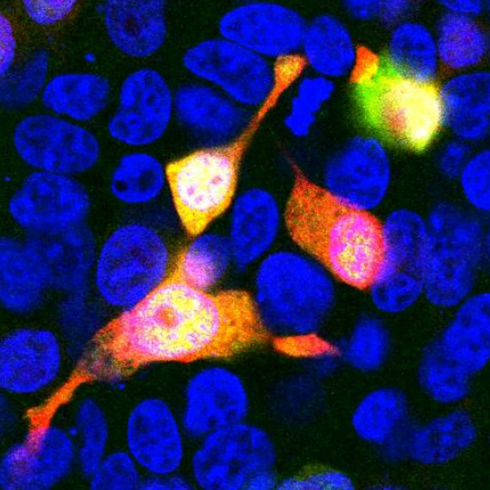
Immunofluorescent analysis of HEK293 cells transfected with mCherry-HA, construct, in red, and stained with mouse mAb to mCherry, dilution 1:500, in green. The blue is Hoechst staining of nuclear DNA. mCherry antibody reveals mCherry protein expressed only in transfected cells which appear golden in color. Untransfected cells do not react with the antibody, as a result only their nuclei are visible.

No documents available.