
MFN1 Rabbit Monoclonal Antibody
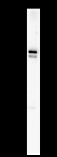
C2C12 whole cell lysates were separated by 10% SDS-PAGE, and the membrane was blotted with MFN1 Rabbit Monoclonal Antibody(1:1000). The HRP-conjugated Goat anti-Rabbit IgG(H + L) antibody was used to detect the antibody.
| Catalog Number: | MABN21056 |
| Conjugate: | Unconjugated |
| Size: | 100 ug |
| Concentration: | 1mg/ml. The concentration of this product may be batch-dependent. |
| Host: | Rabbit |
| Isotype: | IgG,Kappa |
| Reactivity: | Human,Mouse,Rat |
| Applications: | WB 1:2000-1:10000,ICC/IF 1:200-1:1000,ELISA 1:5000-1:20000,IP 1:50-1:200 |
| Molecular Weight: | Calculated MW:84kD;Observed MW:84kD |
| Purification: | Protein A |
| Form: | Liquid |
| Synonyms: | Mitofusin-1;Fzo homolog;Transmembrane GTPase MFN1; |
Cell localization:Mitochondrion outer membrane ; Multi-pass membrane protein .; [Isoform 2]: Cytoplasm ..The protein encoded by this gene is a mediator of mitochondrial fusion. This protein and mitofusin 2 are homologs of the Drosophila protein fuzzy onion (Fzo). They are mitochondrial membrane proteins that interact with each other to facilitate mitochondrial targeting. [provided by RefSeq, Jul 2008]
Applications
WB 1:2000-1:10000,ICC/IF 1:200-1:1000,ELISA 1:5000-1:20000,IP 1:50-1:200