MMP9 Polyclonal Antibody

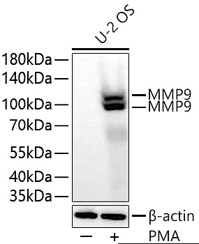
Western blot analysis of lysates from U-2 OS cells usingMMP9 Rabbit pAb at 1:5000 dilution incubatedat room temperature for 1.5 hours. U-2 OS cells were treated with PMA (200 nM) at 37℃ for 48 hours after serumstarvation
overnight.|Secondary antibody: HRP-conjugatedGoat anti-Rabbit IgG (H+L)  at 1:10000 dilution.|Lysates/proteins: 30 μg per lane.| Blocking buffer: 3% nonfatdry milk in TBST.|
Detection: ECL West Pico Plus. 
Exposure time: 20 s.

Western blot analysis of lysates from U-2 OS cells usingMMP9 Rabbit pAb at 1:5000 dilution incubatedat room temperature for 1.5 hours. U-2 OS cells were treated with PMA (200 nM) at 37℃ for 48 hours after serumstarvation overnight.|Secondary antibody: HRP-conjugatedGoat anti-Rabbit IgG (H+L) at 1:10000 dilution.|Lysates/proteins: 30 μg per lane.| Blocking buffer: 3% nonfatdry milk in TBST.| Detection: ECL West Pico Plus. Exposure time: 20 s.

Catalog Number:AB93037
Conjugate:Unconjugated
Size:100 ug
Host:Rabbit
Isotype:IgG
Immunogen:Recombinant fusion protein of human MMP9
Reactivity:Human
Applications:WB: 1:1000-1:5000 IHC 1:50-1:200; IF 1:50-1:200
Molecular Weight:92 kDa / Glycosylated Form: ~106kDa
Purification:Affinity purification
Form:Liquid

Proteins of the matrix metalloproteinase (MMP) family are involved in the breakdown of extracellular matrix in normal physiological processes, such as embryonic development, reproduction, and tissue remodeling, as well as in disease processes, such as arthritis and metastasis. Most MMP's are secreted as inactive proproteins which are activated when cleaved by extracellular proteinases. The enzyme encoded by this gene degrades type IV and V collagens. Studies in rhesus monkeys suggest that the enzyme is involved in IL-8-induced mobilization of hematopoietic progenitor cells from bone marrow, and murine studies suggest a role in tumor-associated tissue remodeling. Depending on the cell type and species (e.g., in mice), glycosylation may cause the bands to migrate slightly higher, between 90kDa and 105 kDa.

Applications

WB: 1:1000-1:5000 IHC 1:50-1:200; IF 1:50-1:200

Immunogen

Recombinant fusion protein of human MMP9
Target Background

Proteins of the matrix metalloproteinase (MMP) family are involved in the breakdown of extracellular matrix in normal physiological processes, such as embryonic development, reproduction, and tissue remodeling, as well as in disease processes, such as arthritis and metastasis. Most MMP's are secreted as inactive proproteins which are activated when cleaved by extracellular proteinases. The enzyme encoded by this gene degrades type IV and V collagens. Studies in rhesus monkeys suggest that the enzyme is involved in IL-8-induced mobilization of hematopoietic progenitor cells from bone marrow, and murine studies suggest a role in tumor-associated tissue remodeling. Depending on the cell type and species (e.g., in mice), glycosylation may cause the bands to migrate slightly higher, between 90kDa and 105 kDa.

Storage

Store at -20°C Valid for 12 months. Avoid freeze / thaw cycles.

Buffer

Phosphate buffered solution, pH 7.4, containing 0.05% stabilizer and 50% glycerol.

Western blot analysis of lysates from U-2 OS cells usingMMP9 Rabbit pAb at 1:5000 dilution incubatedat room temperature for 1.5 hours. U-2 OS cells were treated with PMA (200 nM) at 37℃ for 48 hours after serumstarvation overnight.|Secondary antibody: HRP-conjugatedGoat anti-Rabbit IgG (H+L) at 1:10000 dilution.|Lysates/proteins: 30 μg per lane.| Blocking buffer: 3% nonfatdry milk in TBST.| Detection: ECL West Pico Plus. Exposure time: 20 s.

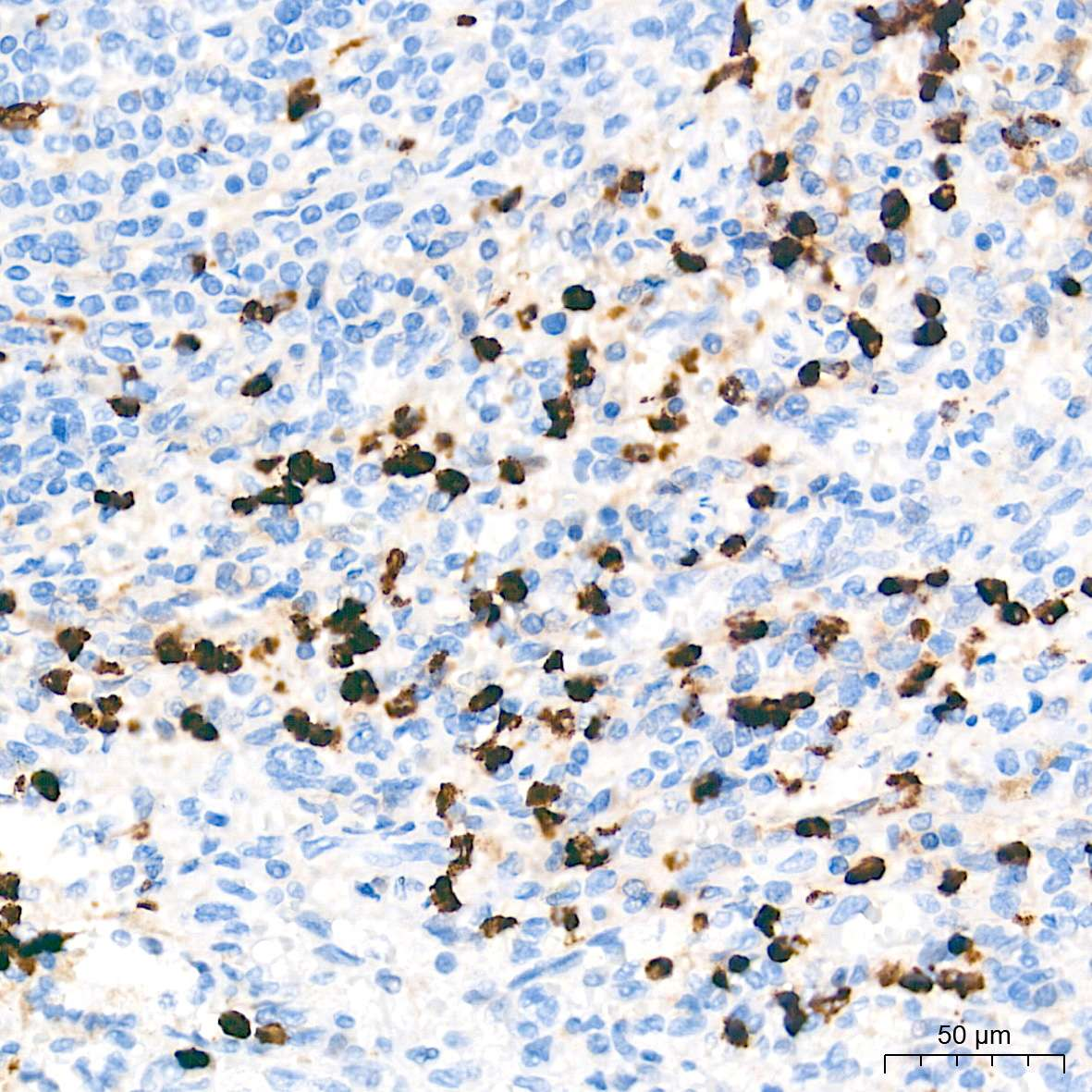
Immunohistochemistry analysis of paraffin-embedded Human spleen tissue using MMP9 Rabbit pAb at a dilution of 1:5000 (40x lens). High pressure antigen retrieval performed with 0.01M Tris-EDTA Buffer (pH 9.0) prior to IHC staining.

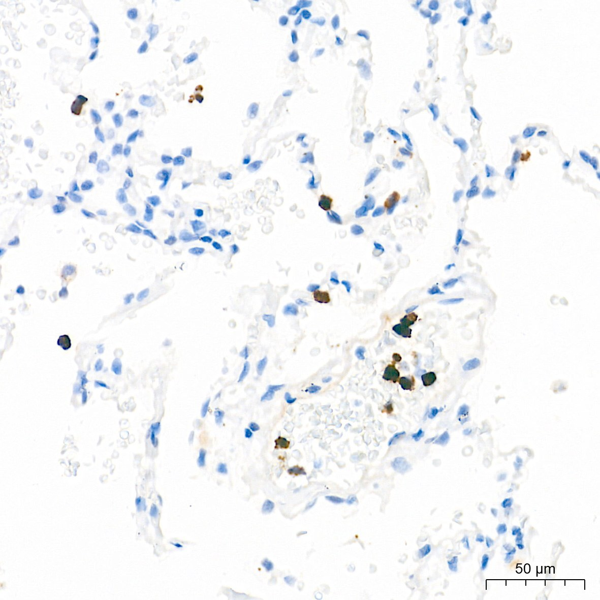
Immunohistochemistry analysis of paraffin-embedded Human lung tissue using MMP9 Rabbit pAb at adilution of 1:5000 (40x lens). High pressure antigen retrievalperformed with 0.01M Tris-EDTA Buffer (pH 9.0) prior to IHC staining.

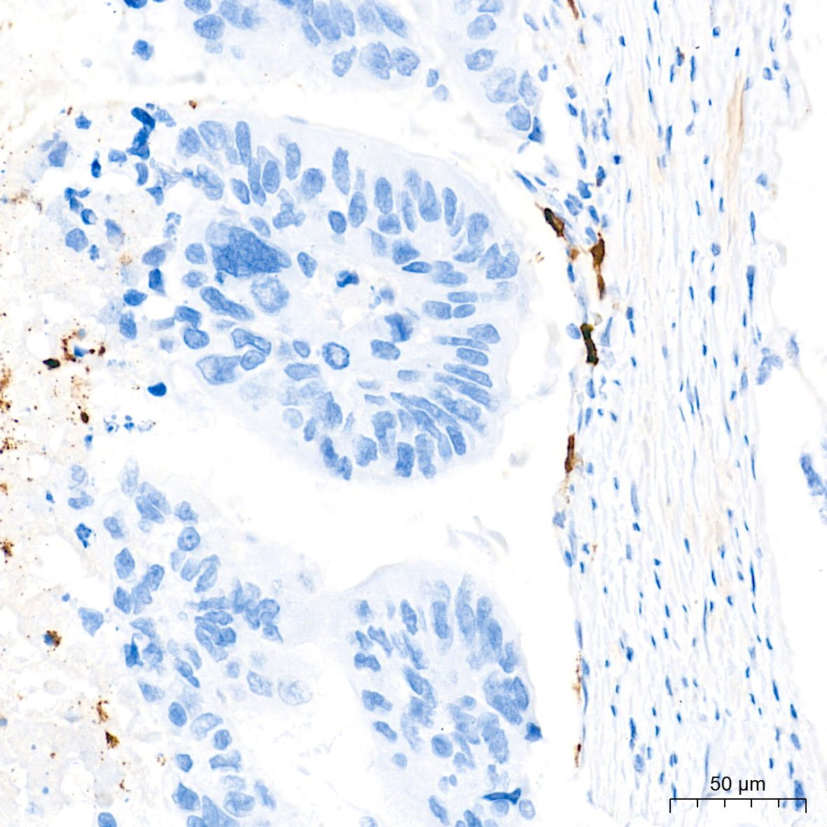
Immunohistochemistry analysis of paraffin-embedded Human colon carcinoma tissue using MMP9 Rabbit pAb at a dilution of 1:5000 (40x lens). High pressure antigen retrieval performed with 0.01M Tris-EDTA Buffer (pH 9.0) prior to IHC staining.

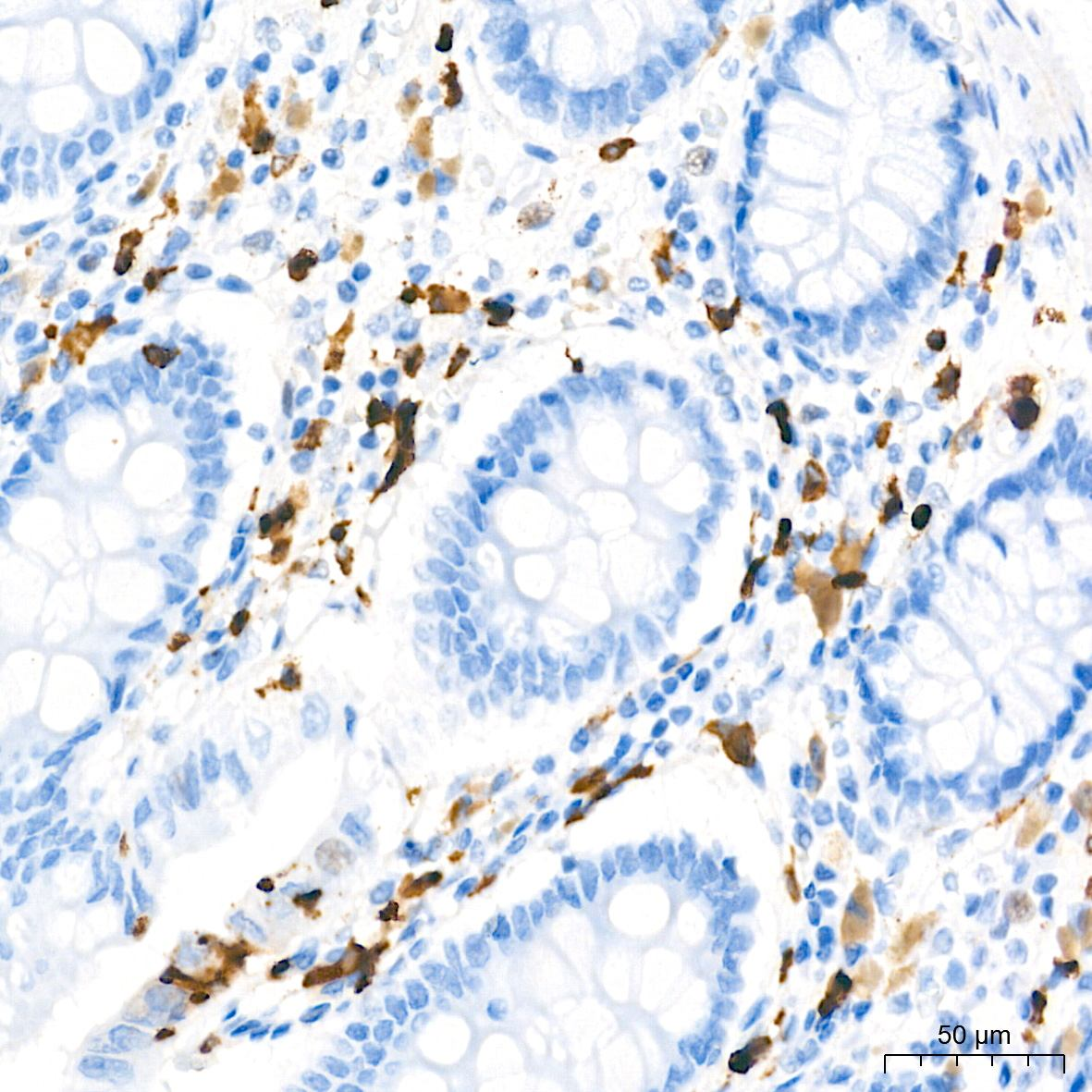
Immunohistochemistry analysis of paraffin-embedded Human colon tissue using MMP9 Rabbit pAb at a dilution of 1:5000 (40x lens). High pressure antigen retrieval performed with 0.01M Tris-EDTA Buffer (pH 9.0) prior to IHC staining.

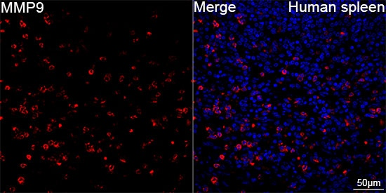
Confocal imaging of paraffin-embedded Human spleen tissue using MMP9 Rabbit pAb (dilution 1:300)followed by a further incubation with Cy3-conjugated Goatanti-Rabbit IgG (H+L) ( dilution 1:500) (Red). DAPI was used for nuclear staining (Blue). High pressure antigen retrieval performed with 0.01M Citrate Buffer (pH 6.0) prior to IF staining. Objective: 40x.

No documents available.