
MUCIN 5AC

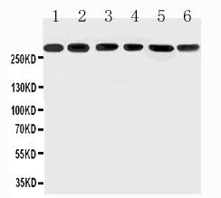
Anti-Mucin 5AC antibody,  All Western blotting
All lanes: Anti-MUC5AC at0.5ug/ml
Lane 1: SGC Whole Cell Lysate at 40ug
Lane 2: HELA Whole Cell Lysate at 40ug
Lane 3: COLO320 Whole Cell Lysate at 40ug
Lane 4: SW620 Whole Cell Lysate at 40ug
Lane 5: MCF-7 Whole Cell Lysate at 40ug
Lane 6: PANC Whole Cell Lysate at 40ug
Predicted bind size: 527KD
Observed bind size: 300KD

Anti-Mucin 5AC antibody, All Western blotting All lanes: Anti-MUC5AC at0.5ug/ml Lane 1: SGC Whole Cell Lysate at 40ug Lane 2: HELA Whole Cell Lysate at 40ug Lane 3: COLO320 Whole Cell Lysate at 40ug Lane 4: SW620 Whole Cell Lysate at 40ug Lane 5: MCF-7 Whole Cell Lysate at 40ug Lane 6: PANC Whole Cell Lysate at 40ug Predicted bind size: 527KD Observed bind size: 300KD

Catalog Number:AB-84263
Size:100ug
Concentration:1mg/ml
Host:Rb
Isotype:IgG
Clone:POLY
Immunogen:A synthetic peptide corresponding to a sequence at the N-terminus of human Mucin 5AC(552-567aa TMQLFMQLAPKLRGQT).
Reactivity:Hu
Applications:Western blot: 0.1-0.5μg/ml Immunohistochemistry(Paraffin-embedded Section) 0.5-1μg/ml
Molecular Weight:585570 MW
Purification:Aff. Pur.
Form:Liquid

Applications

Western blot: 0.1-0.5μg/ml Immunohistochemistry(Paraffin-embedded Section) 0.5-1μg/ml

Immunogen

A synthetic peptide corresponding to a sequence at the N-terminus of human Mucin 5AC(552-567aa TMQLFMQLAPKLRGQT).

Target Background

Mucin-5AC is a protein that in humans is encoded by the MUC5AC gene. It is mapped to 11p15.5. MUC5AC is highly expressed in adult trachea and stomach. This gene has been linked to mucus hypersecretion in the pulmonary tracts and associated to chronic obstructive pulmonary disease(COPD). It has been showed that activation of the NF-kappa-B signaling pathway by inflammatory cytokine TNF-alpha upregulated MUC5AC mRNA expression nearly 6-fold. MUC5AC is also a direct and critical mediator of resistance during intestinal nematode infection.

Storage

At -20°C for one year. It can also be aliquotted and stored frozen at -20°C for a longer time. Avoid repeated freezing and thawing.

Buffer

Each vial contains 5mg BSA, 0.9mg NaCl, 0.2mg Na2HPO4, 0.05mg Thimerosal, 0.05mg NaN3.

Anti-Mucin 5AC antibody, All Western blotting All lanes: Anti-MUC5AC at0.5ug/ml Lane 1: SGC Whole Cell Lysate at 40ug Lane 2: HELA Whole Cell Lysate at 40ug Lane 3: COLO320 Whole Cell Lysate at 40ug Lane 4: SW620 Whole Cell Lysate at 40ug Lane 5: MCF-7 Whole Cell Lysate at 40ug Lane 6: PANC Whole Cell Lysate at 40ug Predicted bind size: 527KD Observed bind size: 300KD

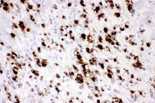
Figure 2. IHC analysis of Mucin 5AC using anti-Mucin 5ACantibody. Mucin 5AC was detected in paraffin-embedded section o fhuman gastric cancer tissue. Heat mediated antigen retrieval was performed in citrate buffer (pH6, epitoperetrieval solution) for 20 mins. The tissue section was blocked with 10% goat serum. The tissue section was then incubated with 1μg/ml rabbit anti-Mucin 5AC Antibody overnight at 4°C. Biotinylated goat anti-rabbit IgG was used as secondary antibody and incubated for 30 minutes at 37°C. The tissue section was developed using Strepavidin-Biotin-Complex (SABC) with DAB as the chromogen.

No documents available.