
Myelin basic protein

Western blot analysis of different tissue lysates using mouse mAb to MBP,dilution 1:10,000 in green: [1] protein standard (red),
[2] rat brain, [3] rat spinal cord, [4] rat sciatic nerve, [5] mouse brain,
[6] mouse spinal cord. Multiple bands at 14kDa, 17kDa, 18.5kDa to
21.5kDa are the alternate transcripts of MBP. Other bands are
proteolytic fragments of the MBP protein.

Western blot analysis of different tissue lysates using mouse mAb to MBP,dilution 1:10,000 in green: [1] protein standard (red), [2] rat brain, [3] rat spinal cord, [4] rat sciatic nerve, [5] mouse brain, [6] mouse spinal cord. Multiple bands at 14kDa, 17kDa, 18.5kDa to 21.5kDa are the alternate transcripts of MBP. Other bands are proteolytic fragments of the MBP protein.

Catalog Number:MAB-94229
Conjugate:Unconjugated
Size:100 ug
Concentration:1mg/ml
Host:Ms
Isotype:IgG1
Clone:7G7
Immunogen:Purified myelin basic protein isolated from bovine brain, epitope is in peptide TPPPSQGKG, amino acids 125-133 of the human 21.5kDa sequence
Reactivity:Hu, Rt, Ms, Cw, Pg, Ho
Applications:Western Blot: 1:5,000 -1:10,000 Immunofluorescence: 1:1,000-2,500 Immunocytochemistry: 1:1,000-2,500 Immunohistochemistry: 1:1,000-2,500
Molecular Weight:14-21.5kDa rodent 17.2-21.5kDa human
Purification:Purified
Form:Liquid

Applications

Western Blot: 1:5,000 -1:10,000 Immunofluorescence: 1:1,000-2,500 Immunocytochemistry: 1:1,000-2,500 Immunohistochemistry: 1:1,000-2,500

Immunogen

Purified myelin basic protein isolated from bovine brain, epitope is in peptide TPPPSQGKG, amino acids 125-133 of the human 21.5kDa sequence

Target Background

Myelin Basic Protein (MBP) is one of the major proteins of the myelin sheath surrounding axons in the nervous system. Since it is of relatively low molecular weight and high abundance the protein sequence was determined from purified protein over 30 years ago (1). The protein is made by oligodendrocytes in the central and nervous system, so antibodies to MBP are good markers of this cell type. In the peripheral nervous system MBP is expressed by myelinating Schwann cells so this antibody can be used to identify these cells in culture or sectioned materials. In the central nervous system four different forms of the protein made by alternate transcription from a single gene, the protein products having molecular weights of 21.5, 20.5, 18.5, and 17.2kDa in humans. The single gene of rodents also produces 4 different proteins but the splicing mechanism is different producing four forms of slightly different sizes, 21.5, 18.5, 17 and 14kDa. Some interest has focused on MBP as a potentially significant auto-antigen involved in mouse models of multiple sclerosis (MS, 3) and in human patients (4). Detection of MBP released into blood and CSF has some potential as a surrogate biomarker of demyelination and axonal loss in MS and other relevant damage and disease states (e.g. 5). The MAB-94229 antibody was made against a preparation of MBP purified biochemically from bovine brain. It can be used to identify oligodendrocytes and Schwann cells in neural cell culture, to visualize myelin sheaths and myelinating cells in sectioned material and to probe western blots for MBP gene products. The antibody is also rather insensitive to aldehyde fixation and so can be used in immunohistochemistry of paraffin sections. The MAB-94229 antibody binds all four of the CNS MBP isoforms, so that the epitope for the antibody is located in the "core" shared by all four gene products. Further mapping localizes the epitope to peptide TPPPSQGKG, amino acids 125-133 of the human 21.5kDa sequence. The data was produced with overlapping peptides which suggests that the last four amino acids, SQGKG, are likely to be key elements of the epitope. This peptide is invariant in rat, mouse, cow and many other species, so this antibody will have wide applicability. In contrast our alternate mouse monoclonal MBP binds only the 21.5kDa and 18.5kDa rat MBP isotypes but all bovine and human isotypes, mapping the epitope toAEGQRPGFGYGGRASDYKSAHKGFKGVDAQGTLSKIFKLG, amino acids 145-184 of the human 21.5kDasequence.

Storage

Store at 4°C for short term, for longer term at -20°C.

Buffer

Purified antibody at 1mg/mL in 50% PBS, 50% glycerol plus 5mM NaN3

Western blot analysis of different tissue lysates using mouse mAb to MBP,dilution 1:10,000 in green: [1] protein standard (red), [2] rat brain, [3] rat spinal cord, [4] rat sciatic nerve, [5] mouse brain, [6] mouse spinal cord. Multiple bands at 14kDa, 17kDa, 18.5kDa to 21.5kDa are the alternate transcripts of MBP. Other bands are proteolytic fragments of the MBP protein.

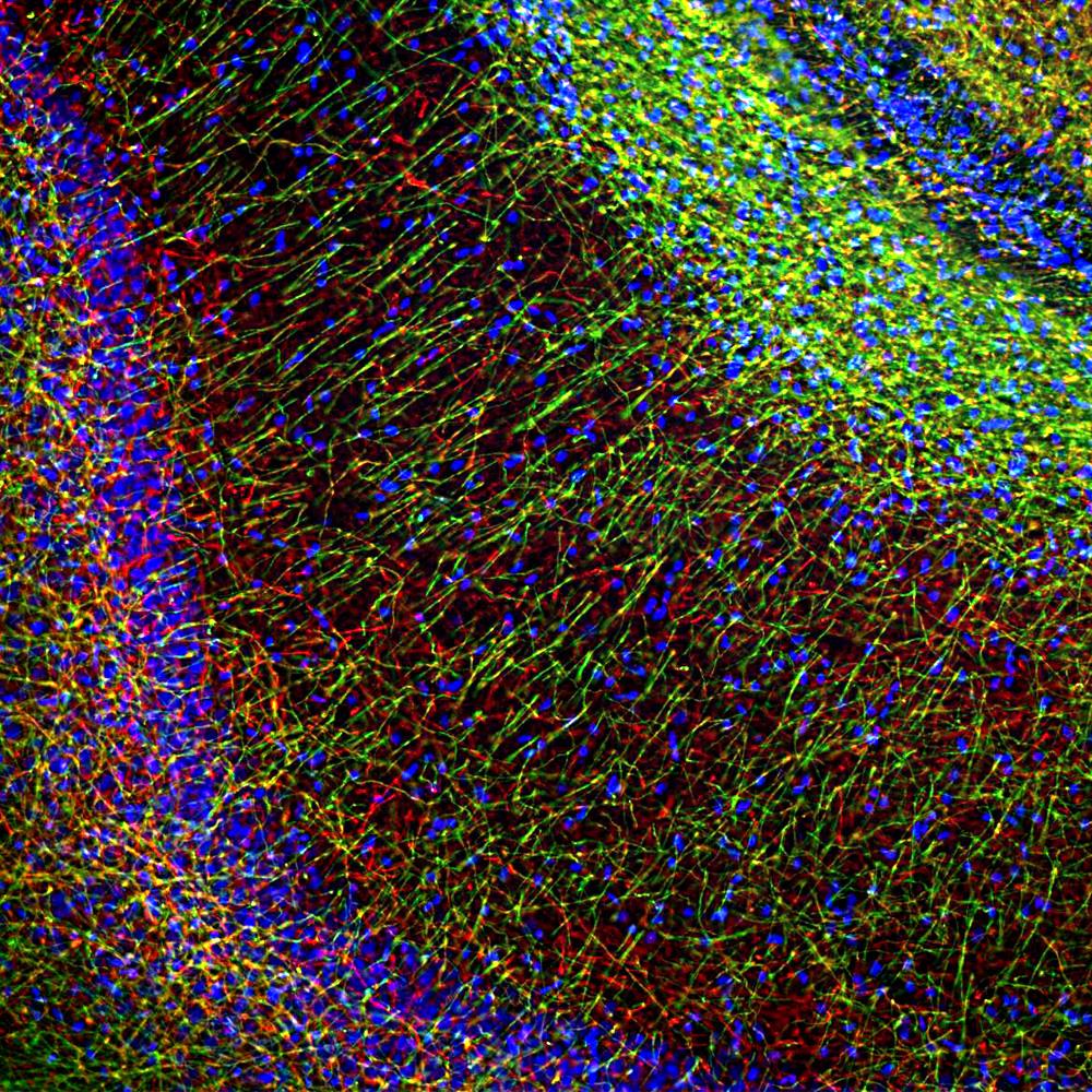
Immunofluorescent analysis of rat hippocampal section stained with mouse mAb to myelin basic protein (MBP), dilution 1:2,500 in green, and costained with rabbit pAb to neurofilament NF-H, dilution 1:2,000, in red. The MBP antibody stains oligodendrocyte cell bodies and the myelin sheathes around axons, while the NF-H antibody labels the axons themselves.

No documents available.