NeuN-FOX3

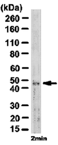
Western Blotting Analysis: Representative lot data. Mouse brain E16 tissue lysate wasprobed with Anti-NeuN (1:500 dilution). Proteins were visualized using a Goat Anti-Guinea Pig IgG secondary antibody conjugated to HRP and a chemiluminescence detection system. Arrow indicates NeuN (~48 kDa)

Western Blotting Analysis: Representative lot data. Mouse brain E16 tissue lysate wasprobed with Anti-NeuN (1:500 dilution). Proteins were visualized using a Goat Anti-Guinea Pig IgG secondary antibody conjugated to HRP and a chemiluminescence detection system. Arrow indicates NeuN (~48 kDa)

Catalog Number:MAB-94161
Conjugate:Unconjugated
Size:500ug
Concentration:1mg/ml
Host:Ms
Isotype:IgG1
Clone:A60
Immunogen:Purified cell nuclei from mouse brain.
Reactivity:Hu, Rt, Ms,Bov, Pr
Applications:WB: 1:100 - 1:1,000 , ICC: 1:100 - 1:200, IHC(F): 1: 100 - 1:1,000, IF, IHC(P): To be determined by end user
Molecular Weight:46-48 kDa
Purification:Purified
Form:Liquid

Applications

WB: 1:100 - 1:1,000 , ICC: 1:100 - 1:200, IHC(F): 1: 100 - 1:1,000, IF, IHC(P): To be determined by end user

Immunogen

Purified cell nuclei from mouse brain.
Target Background

Vertebrate neuron-specific nuclear protein called NeuN (Neuronal Nuclei). Only one NeuN clone exists (A60) and reacts with an uncharacterized nuclear protein. MAB-94161 reacts with most neuronal cell types throughout the nervous system of mice including cerebellum, cerebral cortex, hippocampus, thalamus, spinal cord and neurons in the peripheral nervous system including dorsal root ganglia, sympathetic chain ganglia and enteric ganglia. The immunohistochemical staining is primarily in the nucleus of the neurons with lighter staining in the cytoplasm. The few cell types not reactive with MAB-94161 include Purkinje, mitral and photoreceptor cells. Developmentally, immunoreactivity is first observed shortly after neurons have become postmitotic, no staining has been observed in proliferative zones. The antibody is an excellent marker for neurons in primary cultures and in retinoic acid¬stimulated P19 cells. It is also useful for identifying neurons in transplants.

Storage

Store at 4°C for short term, for longer term at -20°C

Buffer

phosphate buffer, 0.25 M NaCl, pH 7.6 with 0.1% sodium azide

Western Blotting Analysis: Representative lot data. Mouse brain E16 tissue lysate wasprobed with Anti-NeuN (1:500 dilution). Proteins were visualized using a Goat Anti-Guinea Pig IgG secondary antibody conjugated to HRP and a chemiluminescence detection system. Arrow indicates NeuN (~48 kDa)

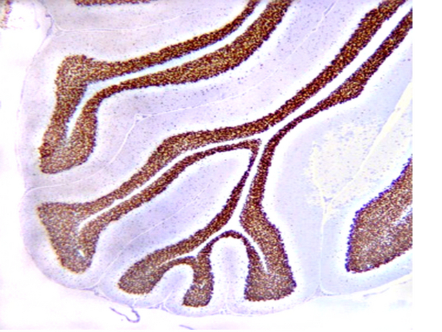
Immunoreactivity in red. Immunohistochemistry(paraffin) Analysis: Optimal Staining With Citrate Buffer, pH 6.0, Epitope Retrieval: Rat Cerebellum NeuN staining pattern/morphology in rat cerebellum. Tissue pretreated with Citrate, pH 6.0. A previous lot of antibody was diluted to 1:100, using IHC-SelectDetection with HRP-DAB. Immunoreactivity is seen as nuclear staining in the neurons in the granular layer.

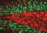
Mouse anti-NeuN and Rabbit anti-Substance P Receptor staining of normal rat hippocampus. NeuN immunoreactivity in green and Substance P Receptor Immunoreactivity in red.

No documents available.