
Neurofilament Heavy (NF-H)

Western blot detection of the heavily phosphorylated axonal form of NFH
protein (pNF-H) in neural tissue lysates (20μg/lane) with affinity
purified mouse monoclonal anti-pNF-H antibody (AH1) at dilution of
1:5,000. Lanes on the blot are: [1] Protein size marker, [2] Adult rat
whole brain [3] Embryonic (E20) rat whole brain.  Adult rat spinal cord
 Adult mouse whole brain.   Adult mouse spinal cord. Rodent pNF-H
protein appears as a single band of about 200kDa in adult rat and
mouse lysates, but is not present in early development (Lane 3).
Additional bands appearing on the blot (Lane 4) are most likely partially
degraded products of pNF-H protein.

Western blot detection of the heavily phosphorylated axonal form of NFH protein (pNF-H) in neural tissue lysates (20μg/lane) with affinity purified mouse monoclonal anti-pNF-H antibody (AH1) at dilution of 1:5,000. Lanes on the blot are: [1] Protein size marker, [2] Adult rat whole brain [3] Embryonic (E20) rat whole brain. Adult rat spinal cord Adult mouse whole brain. Adult mouse spinal cord. Rodent pNF-H protein appears as a single band of about 200kDa in adult rat and mouse lysates, but is not present in early development (Lane 3). Additional bands appearing on the blot (Lane 4) are most likely partially degraded products of pNF-H protein.

Catalog Number:MAB-94404
Conjugate:Unconjugated
Size:100 ug
Concentration:1mg/ml
Host:Ms
Isotype:IgG1
Clone:AH1
Immunogen:Native NF-H purified from bovine spinal cord
Reactivity:Hu Rt Ms,Cw, Pg, horse
Applications:Western Blot: 1:10,000 Immunocytochemistry: 1:1,000 Immunofluorescence: 1:1,000 Immunohistochemistry: 1:1,000
Molecular Weight:200-220 kDa
Purification:Purified
Form:Liquid

Applications

Western Blot: 1:10,000 Immunocytochemistry: 1:1,000 Immunofluorescence: 1:1,000 Immunohistochemistry: 1:1,000

Immunogen

Native NF-H purified from bovine spinal cord
Target Background

Neurofilaments are the 10nm or intermediate filament proteins found specifically in neurons, and are composed predominantly of three major proteins called NF-L, NF-M and NF-H, though other proteins may also be present. NF-H is the neurofilament high or heavy molecular weight polypeptide and runs on SDSPAGE gels at 200-220 kDa, with some variability across species boundaries. The protein is in reality much smaller in molecular size, about 110kDa (1,2). The unusual SDS-PAGE mobility is due partly to a very high content of charged amino acids, particularly glutamic acid rich regions, and the non-phosphorylated form runs on SDS-PAGE at about 160kDa. The predominant type of NF-H is the axonal form which is heavily serine phosphorylated on 40 or more tandemly repeated lysine-serine-proline (KSP) containing peptides (3-5). The phosphorylation of these peptides results in considerable further retardation on SDS-PAGE gels,so the heavily phosphorylated axonal form runs at 200-220kDa with some species variability. Antibodies to NF-H are useful for identifying axonal processes in tissue sections and in culture. NF-H antibodies can also be useful in visualizing neurofilament accumulations seen in many neurological disorders, such as Amyotrophic Lateral Sclerosis (also known as Lou Gehrig’s disease), Alzheimer’s disease and following traumatic injury. The phosphorylated axonal form of NF-H, usually referred to as pNF-H, can be detected in blood and CSF following a variety of damage and disease states resulting in axonal compromise, and antibodies such as this can be used to used to quantify such ongoing axonal loss. MAB-94404 is a mouse monoclonal antibody raised against native axonal phosphorylated NF-H purified from bovine spinal cord (9). MAB-94404 recognizes phosphorylated NF-H KSP sequences but not non phosphorylated KSP sequences, similar to other antibodies to NF-H. In some species there is some cross-reactivity with the phosphorylated KSP sequences found in the related neurofilament subunit NF-M. The antibody recognizes NF-H strongly in all mammals tested to date and also in chicken. It recognizes neurofilaments in frozen sections in tissue culture and in formalin fixed sections.

Storage

Store at 4°C. For long term storage, leave frozen at -20°C

Buffer

Purified antibody at 1mg/mL in 50% PBS, 50% glycerol plus 5mM NaN3

Western blot detection of the heavily phosphorylated axonal form of NFH protein (pNF-H) in neural tissue lysates (20μg/lane) with affinity purified mouse monoclonal anti-pNF-H antibody (AH1) at dilution of 1:5,000. Lanes on the blot are: [1] Protein size marker, [2] Adult rat whole brain [3] Embryonic (E20) rat whole brain. Adult rat spinal cord Adult mouse whole brain. Adult mouse spinal cord. Rodent pNF-H protein appears as a single band of about 200kDa in adult rat and mouse lysates, but is not present in early development (Lane 3). Additional bands appearing on the blot (Lane 4) are most likely partially degraded products of pNF-H protein.

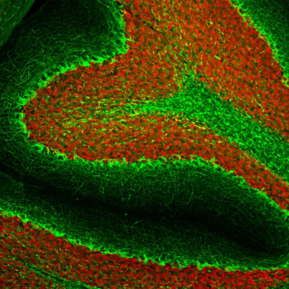
Immunohistological analysis of rat cerebellum section stained with mouse mAb to pNF-H, AH1, dilution 1:2,000 in green, and costained with rabbit pAb to FOX3/NeuN, FOX3, dilution 1:5,000 in red. Following transcardial perfusion with 4% paraformaldehyde, brain was post fixed for 24 hours, cut to 45μM, and free-floating sections were stained with above antibodies. The AH1 antibody stains axons in the granular layer and white matter and prominent basket cell axons surrounding the large Purkinje neurons. The FOX3/NeuN antibody specifically labels nuclei of granular and other neurons, but does not stain Purkinje cells.

No documents available.