NFKB p65

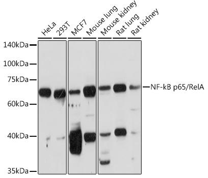
Western blot analysis of extracts of
various cell lines, using NF-kB p65
antibody  at 1:1000 dilution.
Secondary antibody: HRP Goat Anti-Rabbit IgG (H+L) at 1:10000 dilution.
Lysates/proteins: 25ug per lane.
Blocking buffer: 3% nonfat dry milk in TBST.
Detection: ECL West Pico Plus 
Exposure time: 90s.

Western blot analysis of extracts of various cell lines, using NF-kB p65 antibody at 1:1000 dilution. Secondary antibody: HRP Goat Anti-Rabbit IgG (H+L) at 1:10000 dilution. Lysates/proteins: 25ug per lane. Blocking buffer: 3% nonfat dry milk in TBST. Detection: ECL West Pico Plus Exposure time: 90s.

Catalog Number:AB-82033
Conjugate:Unconjugated
Size:100 ug
Concentration:1mg/ml
Host:Rb
Isotype:IgG
Clone:POLY
Immunogen:Recombinant fusion protein containing a sequence corresponding to amino acids 50-180 of human NF-kB p65.
Reactivity:Hu, Ms, Rt
Applications:Western Blot: 1:1000 Immunohistochemistry: 1:50 - 1:100 Immunofluorescence: 1:50 - 1:100 Immunoprecipitation: 1:50 - 1:100
Molecular Weight:65 kDa
Purification:Aff. Pur.
Form:Liquid
Synonyms:RELA; NFKB3; p65; transcription factor p65

Applications

Western Blot: 1:1000 Immunohistochemistry: 1:50 - 1:100 Immunofluorescence: 1:50 - 1:100 Immunoprecipitation: 1:50 - 1:100

Immunogen

Recombinant fusion protein containing a sequence corresponding to amino acids 50-180 of human NF-kB p65.

Target Background

NF-kappa-B is a ubiquitous transcription factor involved in several biological processes. It is held in the cytoplasm in an inactive state by specific inhibitors. Upon degradation of the inhibitor, NF-kappa-B moves to the nucleus and activates transcription of specific genes. NF-kappa-B is composed of NFKB1 or NFKB2 bound to either REL, RELA, or RELB. The most abundant form of NF-kappa-B is NFKB1 complexed with the product of this gene, RELA. Four transcript variants encoding different isoforms have been found for this gene.

Synonyms:RELA; NFKB3; p65; transcription factor p65

Storage

Store at -20℃. Avoid freeze / thaw cycles.

Buffer

PBS with 0.02% sodium azide, 50% glycerol, pH7.3.

Western blot analysis of extracts of various cell lines, using NF-kB p65 antibody at 1:1000 dilution. Secondary antibody: HRP Goat Anti-Rabbit IgG (H+L) at 1:10000 dilution. Lysates/proteins: 25ug per lane. Blocking buffer: 3% nonfat dry milk in TBST. Detection: ECL West Pico Plus Exposure time: 90s.

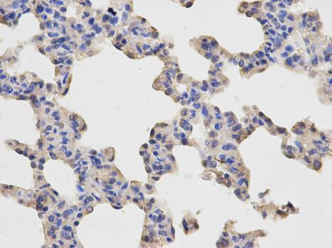
Immunohistochemistry of paraffin-embedded rat lung using NF-kB p65 Antibody at dilution of 1:100 (40x lens).

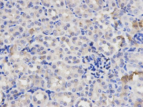
Immunohistochemistry of paraffin-embedded rat kidney using NF-kB p65 Antibody at dilution of 1:100 (40x lens).

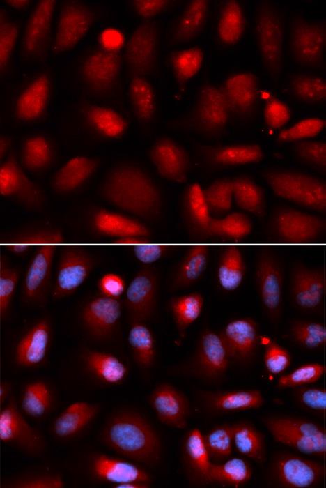
Immunofluorescence analysis of U2OS cells using NF-kB p65 antibody.

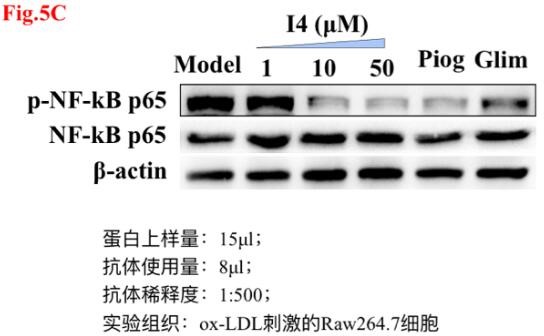
Application: Western blot (WB) Sample: RAW264.7 Loading Amount : 15 ug Dilution: 1:500 Incubation time: over night Temperature 4 ℃ Exposure: 15 seconds Observed Band: 65 kDa Remarks Western blot analysis of extracts of mouse atherosclerosis arterial tissue, using RELA antibody at 1:500 dilution.The band is specific and clear.

No documents available.