NFkB p65 Mouse mAb

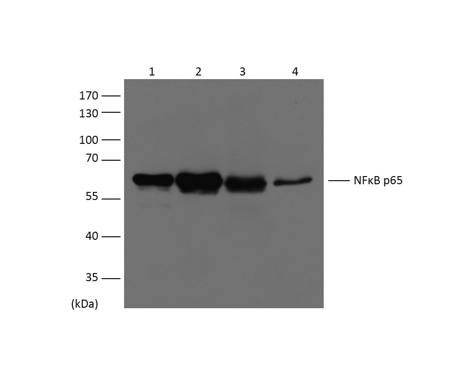
Western blot analysis of extracts from HeLa (Lane 1),MCF-7 (Lane 2), Mouse Liver (Lane 3), Rat Kidney (Lane 4), using NFκB p65 diluted at 1:1,500.

Western blot analysis of extracts from HeLa (Lane 1),MCF-7 (Lane 2), Mouse Liver (Lane 3), Rat Kidney (Lane 4), using NFκB p65 diluted at 1:1,500.

Catalog Number:ABE1111
Conjugate:unconjugated
Size:100 ug
Concentration:1mg/mL
Host:Mouse
Isotype:IgG1
Clone:MB-9
Immunogen:Recombinant Protein
Reactivity:Human, Rat, Mouse
Applications:WB 1:500-2,000 IP 1:200 IF 1:100-200 IHC 1:200-500
Molecular Weight:65kDa
Purification:Affinity Purification
Form:liquid

NFkB p65 is ubiquitinated leading to its proteosomal degradation, which is required for termination of the NFkB response. Phosphorylation of NFkB p65 on S536 stimulates acetylation of K310 by CBP, enhancing transcriptional activity. NFkB p65 is also acetylated at K122, enhancing DNA binding and impairing the interaction with NFKBIA. The protein is deacetylated by HDAC3. Invasion of a host by a pathogen is frequently associated with the activation of NF-kB, which coordinates various aspects of immune function required for resistance to infection.

Applications

WB 1:500-2,000 IP 1:200 IF 1:100-200 IHC 1:200-500

Immunogen

Recombinant Protein
Target Background

NFkB p65 is ubiquitinated leading to its proteosomal degradation, which is required for termination of the NFkB response. Phosphorylation of NFkB p65 on S536 stimulates acetylation of K310 by CBP, enhancing transcriptional activity. NFkB p65 is also acetylated at K122, enhancing DNA binding and impairing the interaction with NFKBIA. The protein is deacetylated by HDAC3. Invasion of a host by a pathogen is frequently associated with the activation of NF-kB, which coordinates various aspects of immune function required for resistance to infection.

Storage

PBS with 0.02% sodium azide and 50% glycerol pH 7.4. Store at -20°C. Avoid repeated freeze-thaw cycles.

Buffer

PBS with 0.02% sodium azide and 50% glycerol pH 7.4

Western blot analysis of extracts from HeLa (Lane 1),MCF-7 (Lane 2), Mouse Liver (Lane 3), Rat Kidney (Lane 4), using NFκB p65 diluted at 1:1,500.

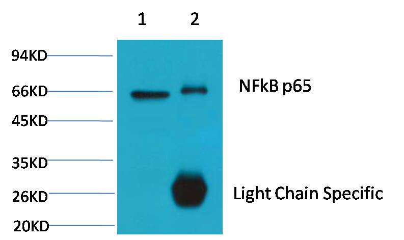
1、Input: Hela Cell Lysate 2、IP product: IP dilute 1:200 Western blot analysis: primary antibody : MAB-94339 1:2,000 Secondary antibody: Goat anti-Mouse IgG, Light chain specific (S003), 1:5,000

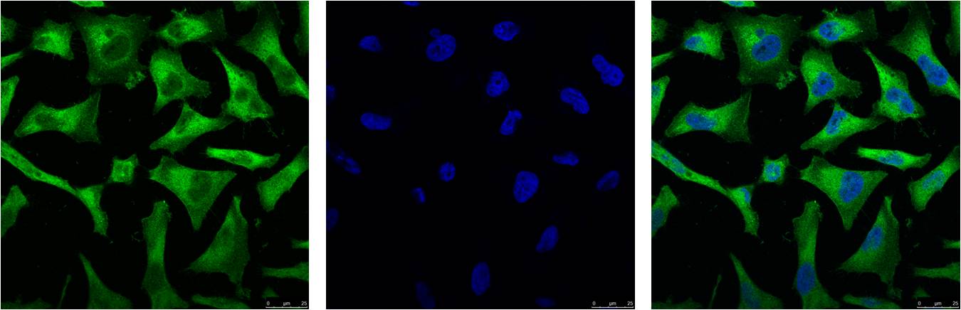
IF analysis of Hela with MAB-94339 (Left) and DAPI (Right) diluted at 1:100.

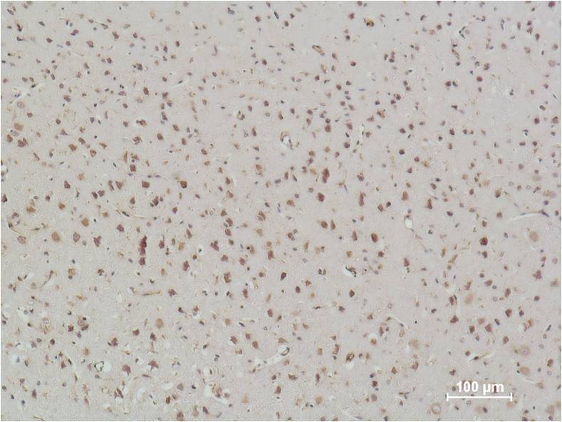
Immunohistochemical analysis of paraffin-embedded Rat Brain Tissue using NFkB p65 MAB-94339 Mouse mAb diluted at 1:500.

No documents available.