
NLRP3

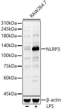
Western blot analysis of RAW264.7, using NLRP3
antibody at 1:770 dilution.Raw264.7 cells
were treated by LPS (1 μg/ml) at 37℃ for 8 hours.
Secondary antibody: HRP Goat Anti-Rabbit IgG (H+L)
at 1:10000 dilution.
Lysates/proteins: 25ug per lane.
Blocking buffer: 3% nonfat dry milk in TBST.
Detection: ECL West Pico Plus.
Exposure time: 60s.

Western blot analysis of RAW264.7, using NLRP3 antibody at 1:770 dilution.Raw264.7 cells were treated by LPS (1 μg/ml) at 37℃ for 8 hours. Secondary antibody: HRP Goat Anti-Rabbit IgG (H+L) at 1:10000 dilution. Lysates/proteins: 25ug per lane. Blocking buffer: 3% nonfat dry milk in TBST. Detection: ECL West Pico Plus. Exposure time: 60s.

Catalog Number:AB-84728
Conjugate:Unconjugated
Size:100 ug
Concentration:1mg/ml
Host:Rb
Isotype:IgG
Clone:POLY
Immunogen:Recombinant fusion protein containing a sequence corresponding to amino acids 1-360 of mouse NLRP3.
Reactivity:Human, Mouse
Applications:Western Blot: 1:500 - 1:2000 Immunofluorescence: 1:50 - 1:200 Immunocytochemistry: 1:50 - 1:200
Molecular Weight:110KDa
Purification:Affinity purification
Form:Liquid
Synonyms:AII; AVP; FCU; MWS; FCAS; KEFH; CIAS1; FCAS1; NALP3; C1orf7; CLR1.1; DFNA34; PYPAF1; AGTAVPRL

Applications

Western Blot: 1:500 - 1:2000 Immunofluorescence: 1:50 - 1:200 Immunocytochemistry: 1:50 - 1:200

Immunogen

Recombinant fusion protein containing a sequence corresponding to amino acids 1-360 of mouse NLRP3.

Target Background

This gene encodes a pyrin-like protein containing a pyrin domain, a nucleotide-binding site (NBS) domain, and a leucine-rich repeat (LRR) motif. This protein interacts with the apoptosis-associated speck-like protein PYCARD/ASC, which contains a caspase recruitment domain, and is a member of the NLRP3 inflammasome complex. This complex functions as an upstream activator of NFkappaB signaling, and it plays a role in the regulation of inflammation, the immune response, and apoptosis. The SARS-CoV 3a protein, a transmembrane pore-forming viroporin, has been shown to activate the NLRP3 inflammasome via the formation of ion channels in macrophages. Mutations in this gene are associated with familial cold autoinflammatory syndrome (FCAS), Muckle-Wells syndrome (MWS), chronic infantile neurological cutaneous and articular (CINCA) syndrome, neonatal-onset multisystem inflammatory disease (NOMID), keratoendotheliitis fugax hereditarian, and deafness, autosomal dominant 34, with or without inflammation. Multiple alternatively spliced transcript variants encoding distinct isoforms have been identified for this gene. Alternative 5' UTR structures are suggested by available data; however, insufficient evidence is available to determine if all of the represented 5' UTR splice patterns are biologically valid.

Synonyms:AII; AVP; FCU; MWS; FCAS; KEFH; CIAS1; FCAS1; NALP3; C1orf7; CLR1.1; DFNA34; PYPAF1; AGTAVPRL

Storage

Store at -20℃. Avoid freeze / thaw cycles.

Buffer

PBS with 0.05% proclin300,50% glycerol,pH7.3.

Western blot analysis of RAW264.7, using NLRP3 antibody at 1:770 dilution.Raw264.7 cells were treated by LPS (1 μg/ml) at 37℃ for 8 hours. Secondary antibody: HRP Goat Anti-Rabbit IgG (H+L) at 1:10000 dilution. Lysates/proteins: 25ug per lane. Blocking buffer: 3% nonfat dry milk in TBST. Detection: ECL West Pico Plus. Exposure time: 60s.

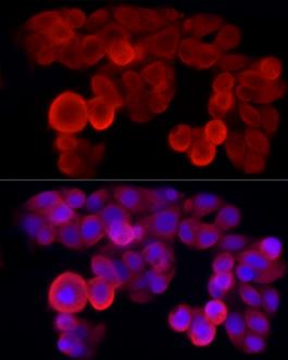
Immunofluorescence analysis of HepG2 cells using NLRP3 Rabbit pAb at dilution of 1:50 (40x lens). Blue: DAPI for nuclear staining.

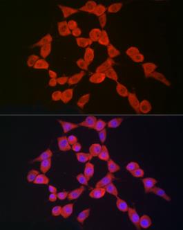
Immunofluorescence analysis of NIH/3T3 cells using NLRP3 Rabbit pAb at dilution of 1:50 (40x lens). Blue: DAPI for nuclear staining

No documents available.