NMDA Receptor 1

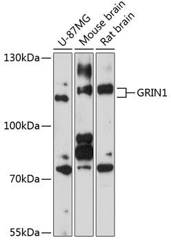
Western blot analysis of extracts of
various cell lines, using GRIN1
antibody at 1:1000 dilution.
Secondary antibody: HRP Goat Anti-
Rabbit IgG (H+L) at 1:10000
dilution.
Lysates/proteins: 25ug per lane.
Blocking buffer: 3% nonfat dry milk in
TBST.
Detection: ECL West Pico Plus.      
Exposure time: 90s.

Western blot analysis of extracts of various cell lines, using GRIN1 antibody at 1:1000 dilution. Secondary antibody: HRP Goat Anti- Rabbit IgG (H+L) at 1:10000 dilution. Lysates/proteins: 25ug per lane. Blocking buffer: 3% nonfat dry milk in TBST. Detection: ECL West Pico Plus. Exposure time: 90s.

Catalog Number:AB-90170
Conjugate:Unconjugated
Size:100 ug
Concentration:1mg/ml
Host:Rb
Isotype:IgG
Clone:POLY
Immunogen:A synthetic peptide corresponding to a sequence within amino acids 600-700 of human GRIN1.
Reactivity:Hu, Ms, Rt
Applications:Western Blot: 1:500 - 1:2000 Immunohistochemistry: 1:50 - 1:200 Immunofluorescence: 1:50 - 1:200
Molecular Weight:110kDa
Purification:Aff. Pur.
Form:Liquid
Synonyms:GRIN1;GluN1;MRD8;NMD-R1;NMDA1;NMDAR1;NR1;NMDA1

Applications

Western Blot: 1:500 - 1:2000 Immunohistochemistry: 1:50 - 1:200 Immunofluorescence: 1:50 - 1:200

Immunogen

A synthetic peptide corresponding to a sequence within amino acids 600-700 of human GRIN1.
Target Background

The protein encoded by this gene is a critical subunit of N-methyl-D-aspartate receptors, members of the glutamate receptor channel superfamily which are heteromeric protein complexes with multiple subunits arranged to form a ligandgated ion channel. These subunits play a key role in the plasticity of synapses, which is believed to underlie memory and learning. Cell-specific factors are thought to control expression of different isoforms, possibly contributing to the functional diversity of the subunits. Alternatively, spliced transcript variants have been described.

Synonyms:GRIN1;GluN1;MRD8;NMD-R1;NMDA1;NMDAR1;NR1;NMDA1

Storage

Store at -20℃. Avoid freeze / thaw cycles.

Buffer

PBS with 0.02% sodium azide,50% glycerol,pH7.3.

Western blot analysis of extracts of various cell lines, using GRIN1 antibody at 1:1000 dilution. Secondary antibody: HRP Goat Anti- Rabbit IgG (H+L) at 1:10000 dilution. Lysates/proteins: 25ug per lane. Blocking buffer: 3% nonfat dry milk in TBST. Detection: ECL West Pico Plus. Exposure time: 90s.

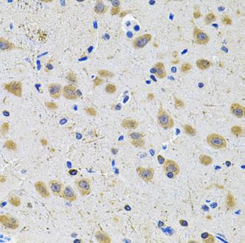
Immunohistochemistry of paraffinembedded rat brain using GRIN1 Antibody at dilution of 1:100 (40x lens).

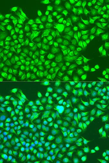
Immunofluorescence analysis of U2OS cells using GRIN1 antibody at dilution of 1:100. Blue: DAPI for nuclear staining.

No documents available.