NOS2(I/NOS) Rabbit Polyclonal Antibody

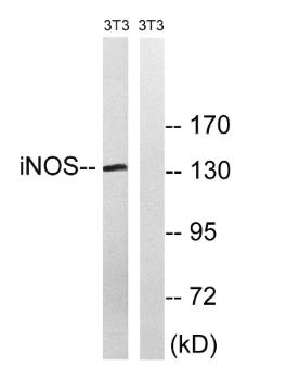
Western blot analysis of lysates from NIH/3T3 cells, using iNOS Antibody.
The lane on the right is blocked with the synthesized peptide.

Western blot analysis of lysates from NIH/3T3 cells, using iNOS Antibody. The lane on the right is blocked with the synthesized peptide.

Catalog Number:AB-80658
Conjugate:Unconjugated
Size:100 ug
Concentration:1mg/ml
Host:Rabbit
Isotype:IgG
Clone:POLY
Immunogen:The antiserum was produced against synthesized peptide derived from human iNOS. AA range:117-166
Reactivity:Hu, Ms, Rt
Applications:Western Blot: 1:500-2000 Immunofluorescence: 1:50-200 Immunohistochemistry: 1:50-300 ELISA: 1:10000-20000
Molecular Weight:131kDa
Purification:The antibody was affinity-purified from rabbit antiserum by affinity-chromatography using epitope-specific immunogen.
Form:Liquid
Synonyms:NOS2; NOS2A; Nitric oxide synthase; inducible; Hepatocyte NOS; HEP-NOS; Inducible NO synthase; Inducible NOS; iNOS; NOS type II; Peptidyl-cysteine S-nitrosylase NOS2

Applications

Western Blot: 1:500-2000 Immunofluorescence: 1:50-200 Immunohistochemistry: 1:50-300 ELISA: 1:10000-20000

Immunogen

The antiserum was produced against synthesized peptide derived from human iNOS. AA range:117-166
Target Background

Nitric oxide is a reactive free radical which acts as a biologic mediator in several processes, including neurotransmission and antimicrobial and antitumoral activities. This gene encodes a nitric oxide synthase which is expressed in liver and is inducible by a combination of lipopolysaccharide and certain cytokines. Three related pseudogenes are located within the Smith-Magenis syndrome region on chromosome 17.

Synonyms:NOS2; NOS2A; Nitric oxide synthase; inducible; Hepatocyte NOS; HEP-NOS; Inducible NO synthase; Inducible NOS; iNOS; NOS type II; Peptidyl-cysteine S-nitrosylase NOS2

Storage

Store at -20°C. Avoid repeated freeze-thaw cycles.

Buffer

Liquid in PBS containing 50% glycerol, 0.5% BSA and 0.02% sodium azide.

Western blot analysis of lysates from NIH/3T3 cells, using iNOS Antibody. The lane on the right is blocked with the synthesized peptide.

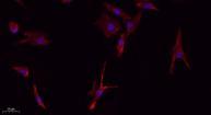
Immunofluorescence analysis of A549. 1,primary Antibody(red) was diluted at 1:200(4°C overnight). 2, Goat Anti Rabbit IgG (H&L) - Alexa Fluor 594 Secondary antibody was diluted at 1:1000(room temperature, 50min). 3, Picture B: DAPI(blue) 10min.

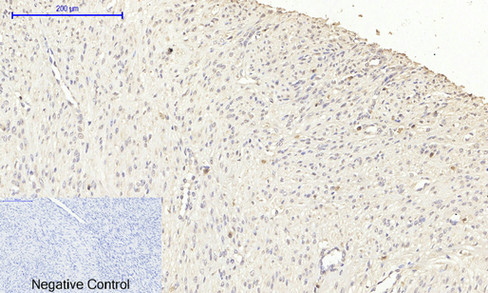
Immunohistochemical analysis of paraffin-embedded Human-uterus-cancer tissue. 1,NOS2 Polyclonal Antibody was diluted at 1:200(4°C,overnight). 2, Sodium citrate pH 6.0 was used for antibody retrieval(>98°C,20min). 3,Secondary antibody was diluted at 1:200(room tempeRature, 30min). Negative control was used by secondary antibody only.

No documents available.