NOTCH 3 Rabbit Polyclonal Antibody

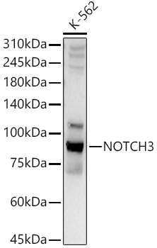
Western blot analysis of lysates from K-562 cells, using NOTCH3 Rabbit pAb at 1:2000 dilution.
Secondary antibody: HRP Goat Anti-Rabbit IgG (H+L) at 1:10000 dilution.
Lysates/proteins: 25μg per lane.
Blocking buffer: 3% nonfat dry milk in TBST.
Detection: ECL West Pico Plus.     
Exposure time: 10s.

Western blot analysis of lysates from K-562 cells, using NOTCH3 Rabbit pAb at 1:2000 dilution. Secondary antibody: HRP Goat Anti-Rabbit IgG (H+L) at 1:10000 dilution. Lysates/proteins: 25μg per lane. Blocking buffer: 3% nonfat dry milk in TBST. Detection: ECL West Pico Plus. Exposure time: 10s.

Catalog Number:AB-10483
Conjugate:Unconjugated
Size:100μg
Concentration:1mg/ml
Host:Rabbit
Isotype:IgG
Clone:POLY
Immunogen:A synthetic peptide corresponding to a sequence within amino acids 2200-2321 of human NOTCH3.
Reactivity:Human,Mouse,Rat
Applications:Western Blot: 1:1000 - 1:5000 Immunohistochemistry(paraffin-embedded tissues): 1:50 - 1:200
Molecular Weight:90kDa
Purification:Affinity purification
Form:Liquid
Synonyms:IMF2; LMNS; CASIL; CADASIL; CADASIL1; NOTCH3

Applications

Western Blot: 1:1000 - 1:5000 Immunohistochemistry(paraffin-embedded tissues): 1:50 - 1:200

Immunogen

A synthetic peptide corresponding to a sequence within amino acids 2200-2321 of human NOTCH3.
Target Background

This gene encodes the third discovered human homologue of the Drosophilia melanogaster type I membrane protein notch. In Drosophilia, notch interaction with its cell-bound ligands (delta, serrate) establishes an intercellular signalling pathway that plays a key role in neural development. Homologues of the notch-ligands have also been identified in human, but precise interactions between these ligands and the human notch homologues remains to be determined. Mutations in NOTCH3 have been identified as the underlying cause of cerebral autosomal dominant arteriopathy with subcortical infarcts and leukoencephalopathy (CADASIL).

Synonyms:IMF2; LMNS; CASIL; CADASIL; CADASIL1; NOTCH3

Storage

Store at -20℃. Avoid freeze / thaw cycles.

Buffer

PBS with 0.02% sodium azide,50% glycerol,pH7.3.

Western blot analysis of lysates from K-562 cells, using NOTCH3 Rabbit pAb at 1:2000 dilution. Secondary antibody: HRP Goat Anti-Rabbit IgG (H+L) at 1:10000 dilution. Lysates/proteins: 25μg per lane. Blocking buffer: 3% nonfat dry milk in TBST. Detection: ECL West Pico Plus. Exposure time: 10s.

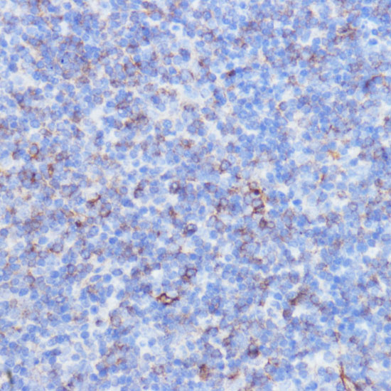
Immunohistochemistry analysis of paraffin-embedded mouse spleen using NOTCH3 Rabbit pAb at dilution of 1:100 (40x lens).Perform microwave antigen retrieval with 10 mM PBS buffer pH 7.2 before commencing with IHC staining protocol.

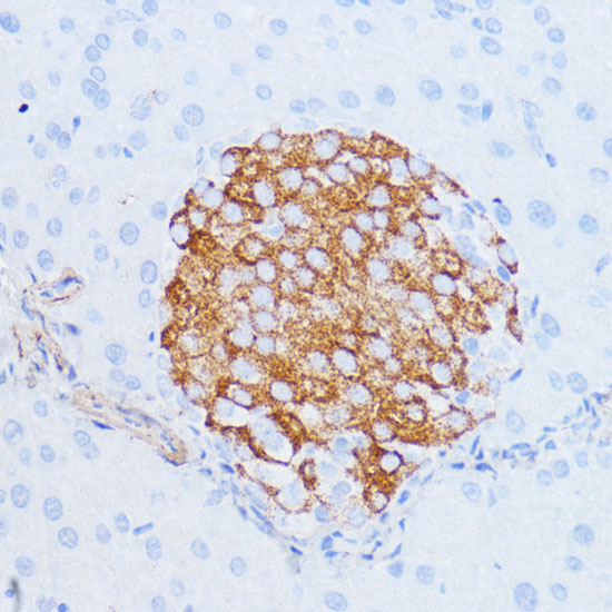
Immunohistochemistry analysis of paraffin-embedded mouse pancreas using NOTCH3 Rabbit pAb at dilution of 1:100 (40x lens).Perform microwave antigen retrieval with 10 mM PBS buffer pH 7.2 before commencing with IHC staining protocol.

No documents available.