NRF1

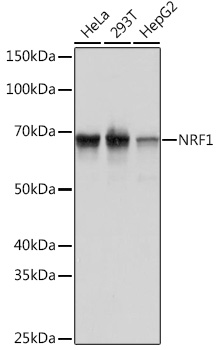
Western blot analysis of extracts of various cell lines, using NRF1 Rabbit mAb at 1:1000 dilution.
Secondary antibody: HRP Goat Anti-Rabbit IgG (H+L) at 1:10000 dilution.
Lysates/proteins: 25ug per lane.
Blocking buffer: 3% nonfat dry milk in TBST.
Detection: ECL Basic Kit.
Exposure time: 1s.

Western blot analysis of extracts of various cell lines, using NRF1 Rabbit mAb at 1:1000 dilution. Secondary antibody: HRP Goat Anti-Rabbit IgG (H+L) at 1:10000 dilution. Lysates/proteins: 25ug per lane. Blocking buffer: 3% nonfat dry milk in TBST. Detection: ECL Basic Kit. Exposure time: 1s.

Catalog Number:AB-81672
Size:100 ug
Concentration:1mg/ml
Host:Rabbit
Isotype:IgG
Clone:POLY
Immunogen:A synthetic peptide corresponding to a sequence within amino acids 300-400 of human NRF1
Reactivity:Human, Mouse, Rat
Applications:Western Blot 1:500 - 1:2000 Immunohistochemistry (P) 1:50 - 1:200 Immunoprecipitation 1:500 - 1:1000
Molecular Weight:68 kDa
Purification:Affinity Purified
Form:Liquid
Synonyms:ALPHA-PAL

Applications

Western Blot 1:500 - 1:2000 Immunohistochemistry (P) 1:50 - 1:200 Immunoprecipitation 1:500 - 1:1000

Immunogen

A synthetic peptide corresponding to a sequence within amino acids 300-400 of human NRF1
Target Background

This gene encodes a protein that homodimerizes and functions as a transcription factor which activates the expression of some key metabolic genes regulating cellular growth and nuclear genes required for respiration, heme biosynthesis, and mitochondrial DNA transcription and replication. The protein has also been associated with the regulation of neurite outgrowth. Alternative splicing results in multiple transcript variants. Confusion has occurred in bibliographic databases due to the shared symbol of NRF1 for this gene and for "nuclear factor (erythroid-derived 2)-like 1" which has an official symbol of NFE2L1.

Synonyms:ALPHA-PAL

Storage

Store at -20℃. Avoid freeze / thaw cycles

Buffer

PBS with 0.02% sodium azide,0.05% BSA,50% glycerol, pH7.3.

Western blot analysis of extracts of various cell lines, using NRF1 Rabbit mAb at 1:1000 dilution. Secondary antibody: HRP Goat Anti-Rabbit IgG (H+L) at 1:10000 dilution. Lysates/proteins: 25ug per lane. Blocking buffer: 3% nonfat dry milk in TBST. Detection: ECL Basic Kit. Exposure time: 1s.

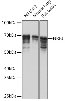
Western blot analysis of extracts of various cell lines, using NRF1 Rabbit mAb at 1:1000 dilution. Secondary antibody: HRP Goat Anti-Rabbit IgG (H+L) at 1:10000 dilution. Lysates/proteins: 25ug per lane. Blocking buffer: 3% nonfat dry milk in TBST. Detection: ECL Basic Kit. Exposure time: 5s.

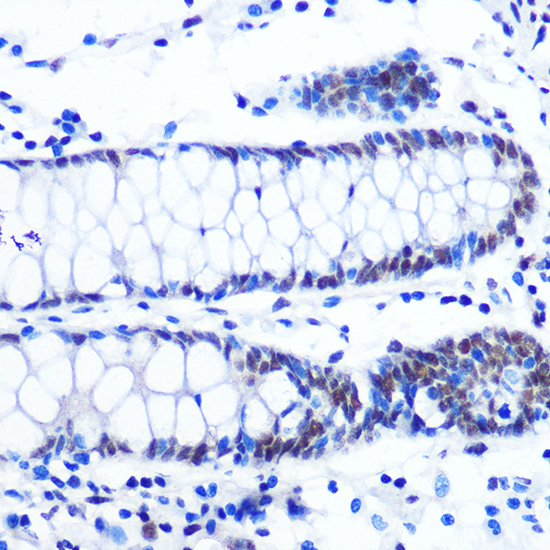
Immunohistochemistry of paraffinembedded human colon using NRF1 Rabbit mAb at dilution of 1:100 (40x lens). Perform microwave antigen retrieval with 10 mM PBS buffer pH 7.2 before commencing with IHC staining protocol.

No documents available.