Oct-4

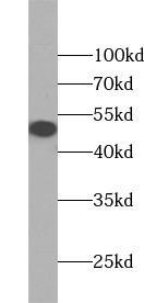
HEK-293 cells were subjected to SDS PAGE followed by western blot with ( OCT4 Antibody) at dilution of 1:1000

HEK-293 cells were subjected to SDS PAGE followed by western blot with ( OCT4 Antibody) at dilution of 1:1000

Catalog Number:AB-84337
Conjugate:Unconjugated
Size:100 ug
Concentration:1mg/ml
Host:Rb
Isotype:IgG
Clone:POLY
Immunogen:POU class 5 homeobox 1
Reactivity:Hu,Ms,Rt
Applications:Western Blot: 1:500 - 1:2000 Immunohistochemistry: 1:50 - 1:200 Immunofuorescence: 1:50 - 1:200
Molecular Weight:50kDa
Purification:Aff. Pur. ≥95% as determined by SDS-PAGE
Form:Liquid
Synonyms:Oct 3, Oct 4, OCT3, OCT3/4, OCT4, Octamer binding protein 3, Octamer binding protein 4, OTF 3, OTF3, OTF4, POU class 5 homeobox 1, POU5F1

Applications

Western Blot: 1:500 - 1:2000 Immunohistochemistry: 1:50 - 1:200 Immunofuorescence: 1:50 - 1:200

Immunogen

POU class 5 homeobox 1
Target Background

This gene encodes a transcription factor containing a POU homeodomain that plays a key role in embryonic development and stem cell pluripotency. Aberrant expression of this gene in adult tissues is associated with tumorigenesis. This gene can participate in a translocation with the Ewing's sarcoma gene on chromosome 21, which also leads to tumor formation. Alternative splicing, as well as usage of alternative AUG and non-AUG translation initiation codons, results in multiple isoforms. One of the AUG start codons is polymorphic in human populations. Related pseudogenes have been identified on chromosomes 1, 3, 8, 10, and 12.

Synonyms:Oct 3, Oct 4, OCT3, OCT3/4, OCT4, Octamer binding protein 3, Octamer binding protein 4, OTF 3, OTF3, OTF4, POU class 5 homeobox 1, POU5F1

Storage

T -20℃ for 12 months (Avoid repeated freeze / thaw cycles.)

Buffer

PBS with 0.02% sodium azide and 50% glycerol pH 7.3

HEK-293 cells were subjected to SDS PAGE followed by western blot with ( OCT4 Antibody) at dilution of 1:1000

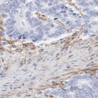
Immunohistochemistry of paraffin-embedded human colon cancer using (OCT4 antibody) at dilution of 1:50

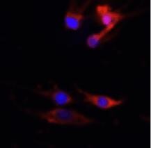
Experiment Type: Immunofluorescence (IF) Sample: HeLa cells Description: Immunofluorescence analysis of HeLa cells using antibody at 1:50 dilution. The positive signal is Golgi staining

No documents available.