Olig2 Rabbit mAb

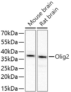
Western blot analysis of lysates from Rat brain using Olig2 Rabbit mAb (MAB26746) at 1:2000 dilution incubated overnight at 4℃.
Secondary antibody: HRP-conjugated Goat anti-Rabbit IgG (H+L) at 1:10000 dilution.
Lysates/proteins: 25 μg per lane.
Blocking buffer: 3% nonfat dry milk in TBST.
Detection: ECL Basic Kit.
Exposure time: 45s.

Western blot analysis of lysates from Rat brain using Olig2 Rabbit mAb (MAB26746) at 1:2000 dilution incubated overnight at 4℃. Secondary antibody: HRP-conjugated Goat anti-Rabbit IgG (H+L) at 1:10000 dilution. Lysates/proteins: 25 μg per lane. Blocking buffer: 3% nonfat dry milk in TBST. Detection: ECL Basic Kit. Exposure time: 45s.

Catalog Number:MAB26746
Conjugate:Unconjugated
Size:100 ug
Concentration:1mg/ml
Host:Rabbit
Isotype:IgG
Immunogen:Synthetic peptide. This information is considered to be commercially sensitive.
Reactivity:Human,Mouse,Rat
Applications:WB 1:1000 - 1:4000 IF-P 1:200 - 1:1000 IHC-P 1:200 - 1:800 ELISA Recommended starting concentration is 1 μg/mL. Please optimize the concentration based on your specific assay requirements.
Molecular Weight:35kDa
Purification:Affinity purification
Form:liquid
Synonyms:BHLHB1; OLIGO2; RACK17; PRKCBP2; bHLHe19

This gene encodes a basic helix-loop-helix transcription factor which is expressed in oligodendroglial tumors of the brain. The protein is an essential regulator of ventral neuroectodermal progenitor cell fate. The gene is involved in a chromosomal translocation t(14;21)(q11.2;q22) associated with T-cell acute lymphoblastic leukemia. Its chromosomal location is within a region of chromosome 21 which has been suggested to play a role in learning deficits associated with Down syndrome.

Applications

WB 1:1000 - 1:4000 IF-P 1:200 - 1:1000 IHC-P 1:200 - 1:800 ELISA Recommended starting concentration is 1 μg/mL. Please optimize the concentration based on your specific assay requirements.

Immunogen

Synthetic peptide. This information is considered to be commercially sensitive.
Target Background

This gene encodes a basic helix-loop-helix transcription factor which is expressed in oligodendroglial tumors of the brain. The protein is an essential regulator of ventral neuroectodermal progenitor cell fate. The gene is involved in a chromosomal translocation t(14;21)(q11.2;q22) associated with T-cell acute lymphoblastic leukemia. Its chromosomal location is within a region of chromosome 21 which has been suggested to play a role in learning deficits associated with Down syndrome.

Synonyms:BHLHB1; OLIGO2; RACK17; PRKCBP2; bHLHe19

Storage

Store at -20℃. Avoid freeze / thaw cycles.

Buffer

PBS with 0.05% BSA,40% glycerol,pH7.3.

Western blot analysis of lysates from Rat brain using Olig2 Rabbit mAb (MAB26746) at 1:2000 dilution incubated overnight at 4℃. Secondary antibody: HRP-conjugated Goat anti-Rabbit IgG (H+L) at 1:10000 dilution. Lysates/proteins: 25 μg per lane. Blocking buffer: 3% nonfat dry milk in TBST. Detection: ECL Basic Kit. Exposure time: 45s.

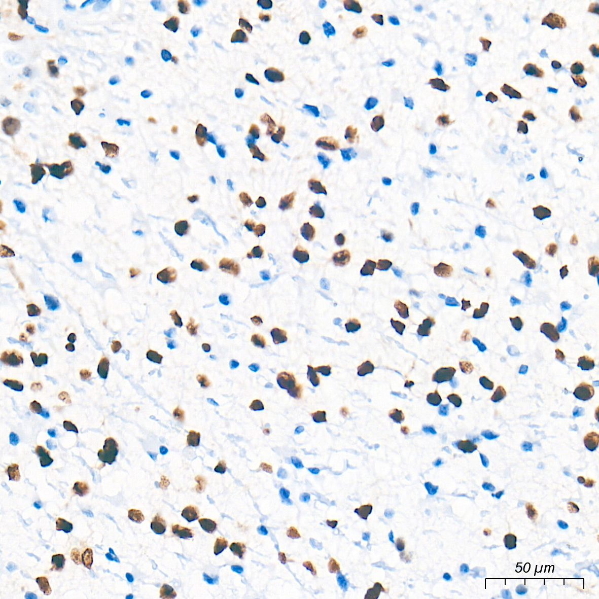
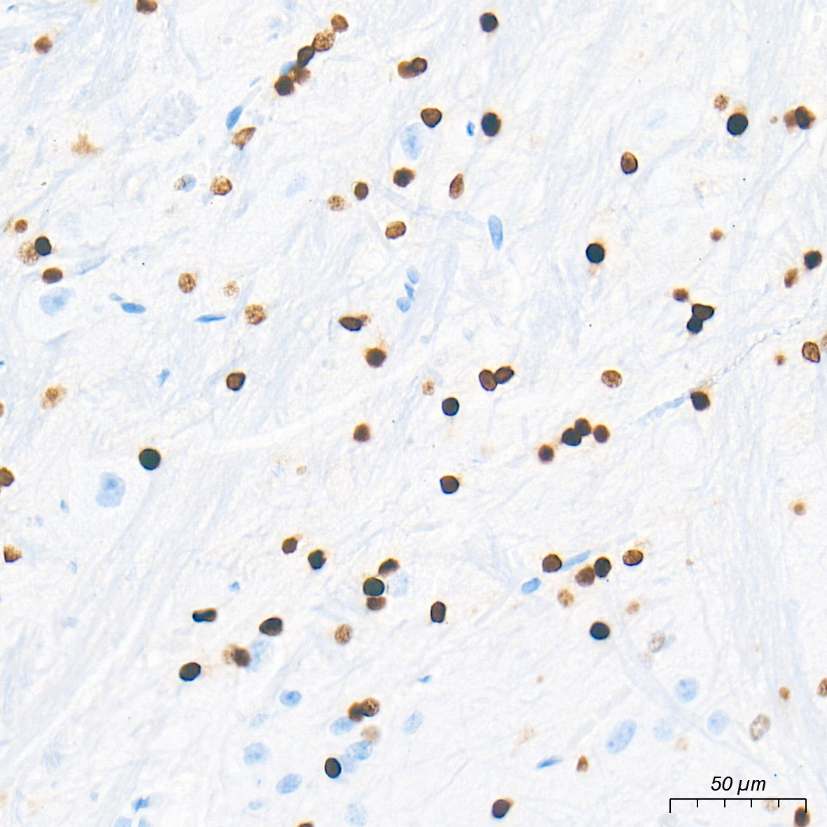
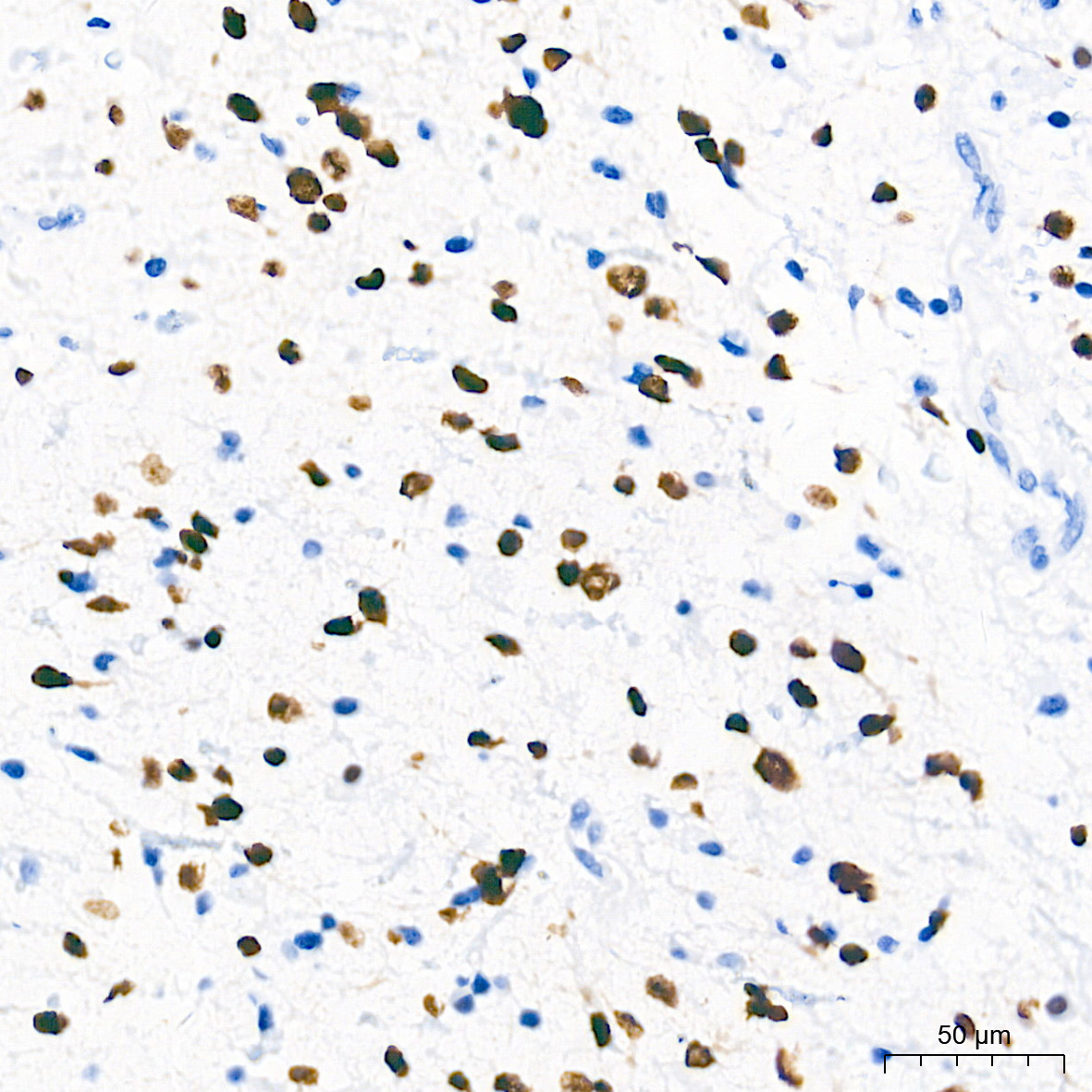
Immunohistochemistry analysis of paraffin-embedded Human brain astrocytoma tissue using Olig2 Rabbit mAb (MAB26746) at a dilution of 1:200 (40x lens). High pressure antigen retrieval performed with 0.01M Tris-EDTA Buffer (pH 9.0) prior to IHC staining.

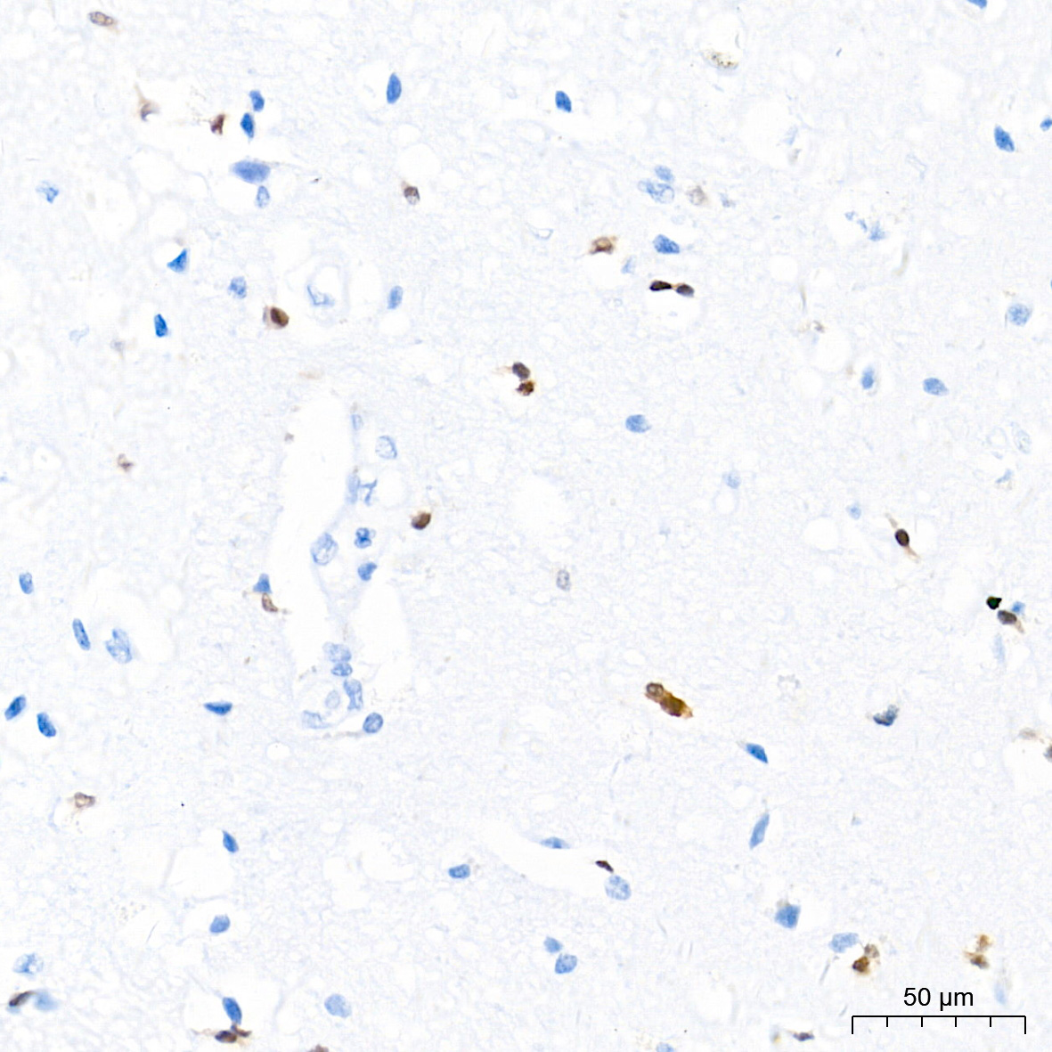
Immunohistochemistry analysis of paraffin-embedded Human brain tissue using Olig2 Rabbit mAb (MAB26746) at a dilution of 1:200 (40x lens). High pressure antigen retrieval performed with 0.01M Tris-EDTA Buffer (pH 9.0) prior to IHC staining.

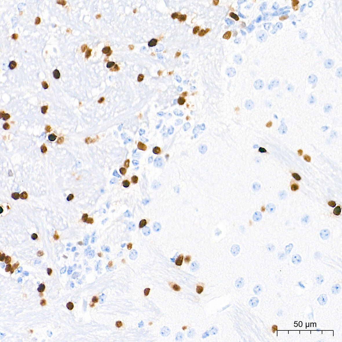
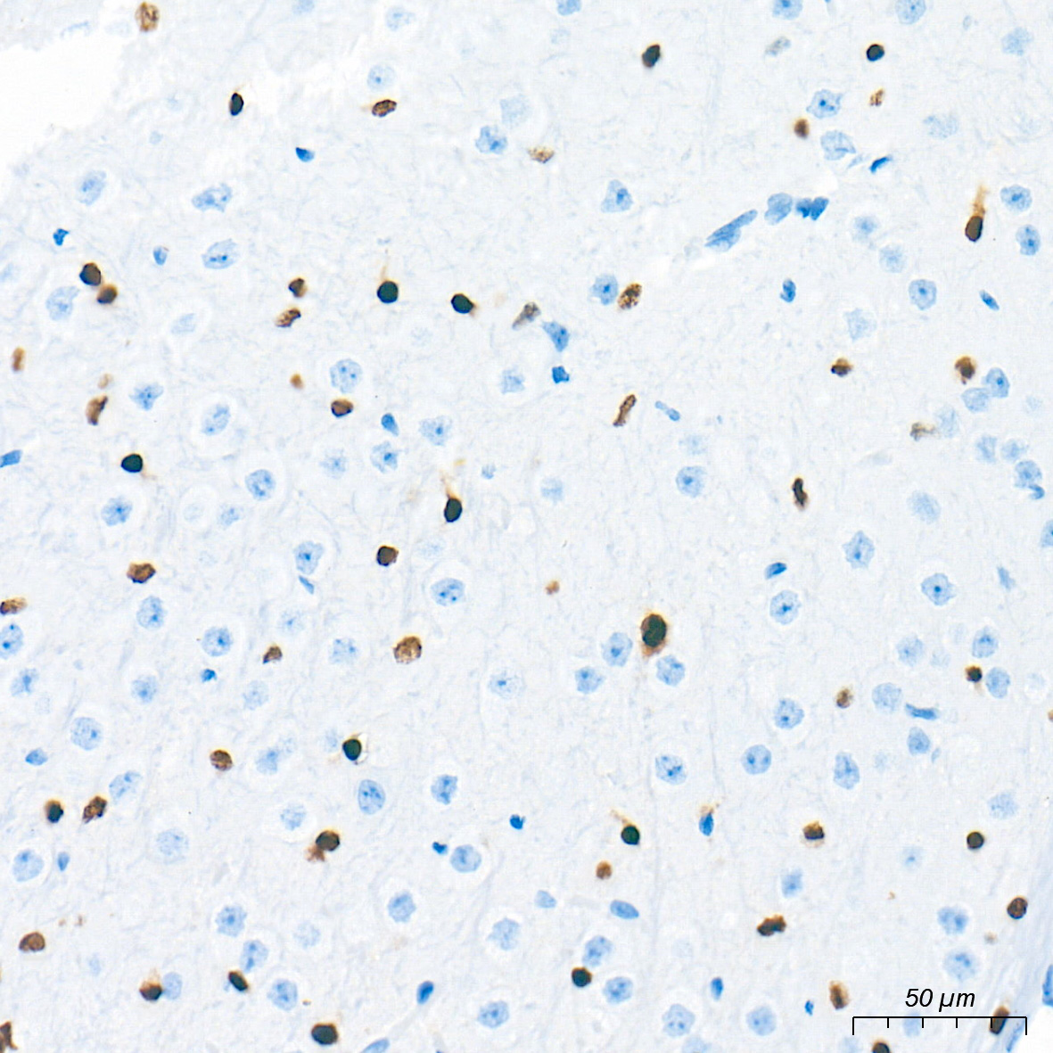
Immunohistochemistry analysis of paraffin-embedded Mouse brain tissue using Olig2 Rabbit mAb (MAB26746) at a dilution of 1:200 (40x lens). High pressure antigen retrieval performed with 0.01M Tris-EDTA Buffer (pH 9.0) prior to IHC staining.

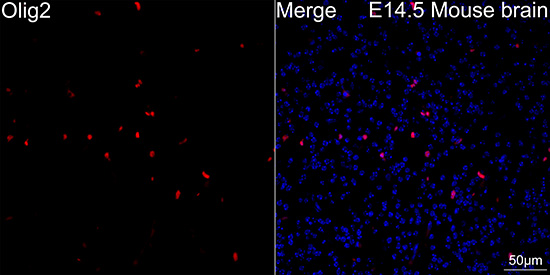
Confocal imaging of paraffin-embedded E14.5 Mouse brain tissue using Olig2 Rabbit mAb (MAB26746, dilution 1:200) followed by a further incubation with Cy3 Goat Anti-Rabbit IgG (H+L) (dilution 1:500) (Red). DAPI was used for nuclear staining (Blue). Microwave antigen retrieval performed with 0.01M Citrate Buffer(pH 6.0) prior to IF staining. Objective: 40x.

Confocal imaging of paraffin-embedded Mouse brain tissue using Olig2 Rabbit mAb (MAB26746, dilution 1:200) followed by a further incubation with Cy3 Goat Anti-Rabbit IgG (H+L) (dilution 1:500) (Red). DAPI was used for nuclear staining (Blue). Microwave antigen retrieval performed with 0.01M Citrate Buffer(pH 6.0) prior to IF staining. Objective: 40x.

Confocal imaging of paraffin-embedded Rat brain tissue using Olig2 Rabbit mAb (MAB26746, dilution 1:200) followed by a further incubation with Cy3 Goat Anti-Rabbit IgG (H+L) (dilution 1:500) (Red). DAPI was used for nuclear staining (Blue). Microwave antigen retrieval performed with 0.01M Citrate Buffer(pH 6.0) prior to IF staining. Objective: 40x.

Confocal imaging of paraffin-embedded Human brain tissue using Olig2 Rabbit mAb (MAB26746, dilution 1:200) followed by a further incubation with Cy3 Goat Anti-Rabbit IgG (H+L) (dilution 1:500) (Red). DAPI was used for nuclear staining (Blue). Microwave antigen retrieval performed with 0.01M Citrate Buffer(pH 6.0) prior to IF staining. Objective: 40x.

No documents available.