OPG Rabbit Polyclonal Antibody

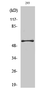
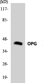
Western Blot analysis of various cells using OPG Polyclonal Antibody diluted at 1:1000

Western Blot analysis of various cells using OPG Polyclonal Antibody diluted at 1:1000

Catalog Number:AB-E11701
Conjugate:Unconjated
Size:100 ug
Concentration:1mg/ml
Host:Rabbit
Isotype:IgG
Clone:POLY
Immunogen:The antiserum was produced against synthesizedpeptide derived from human TR11B . AA range:10-59
Reactivity:Human;Mouse
Applications:Western Blot: 1/500 - 1/2000.Immunohistochemistry(paraffin-embedded tissues): 1:100-300
Molecular Weight:55kD
Purification:The antibody was affinity-purified fromrabbit antiserum by affinity-chromatography using epitope-specific immunogen.
Form:Liquid
Synonyms:TNFRSF11B; OCIF; OPG; Tumor necrosis factor receptor superfamily member 11B; Osteoclastogenesis inhibitory factor; Osteoprotegerin

Applications

Western Blot: 1/500 - 1/2000.Immunohistochemistry(paraffin-embedded tissues): 1:100-300

Immunogen

The antiserum was produced against synthesizedpeptide derived from human TR11B . AA range:10-59
Target Background

The protein encoded by this gene is a member of the TNF-receptor superfamily. This protein is an osteoblast-secreted decoy receptor that functions as a negative regulator of bone resorption. This proteinspecifically binds to its ligand, osteoprotegerin ligand, both of which are key extracellular regulators of osteoclast development. Studies of the mouse counterpart also suggest that this protein and its ligand play a role in lymph-node organogenesis andvascular calcification. Alternatively spliced transcript variants of this gene have been reported, but their full length nature has not been determined.

Synonyms:TNFRSF11B; OCIF; OPG; Tumor necrosis factor receptor superfamily member 11B; Osteoclastogenesis inhibitory factor; Osteoprotegerin

Storage

Store at -20°C. Avoid repeated freeze-thawcycles.

Buffer

Liquid in PBS containing 50% glycerol, 0.5%BSAand0.02% sodium azide.

Western Blot analysis of various cells using OPG Polyclonal Antibody diluted at 1:1000

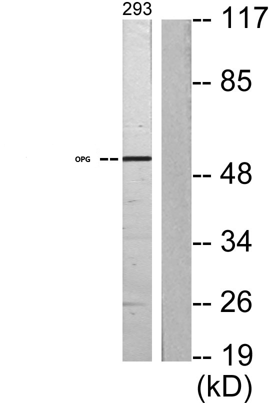
Western blot analysis of lysates from 293 cells, using OPG Antibody . The lane on the right is blocked with the synthesized peptide.

Western blot analysis of the lysates from HeLa cells using OPG antibody

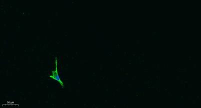
Immunofluorescence analysis of A549. 1,primary Antibody was diluted at 1:200(4°C overnight). 2, Goat Anti Rabbit IgG (H&L) - Alexa Fluor 488 Secondary antibody was diluted at 1:1000(room temperature, 50min).3, Picture B: DAPI(blue) 10min.

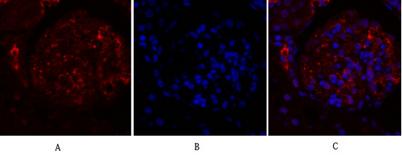
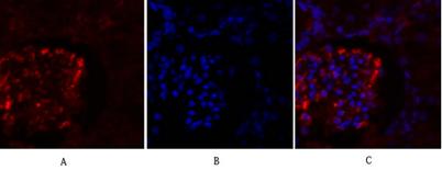
Immunofluorescence analysis of rat-kidney tissue. 1,OPGPolyclonal Antibody(red) was diluted at 1:200(4°C,overnight). 2, Cy3 labled Secondary antibody was diluted at 1:300(room temperature, 50min).3, Picture B: DAPI(blue) 10min. Picture A:Target. Picture B: DAPI. Picture C: merge of A+B

Immunofluorescence analysis of rat-kidney tissue. 1,OPG Polyclonal Antibody(red) was diluted at 1:200(4°C,overnight). 2, Cy3 labled Secondary antibody was diluted at 1:300(room temperature, 50min).3, Picture B: DAPI(blue) 10min. Picture A:Target. Picture

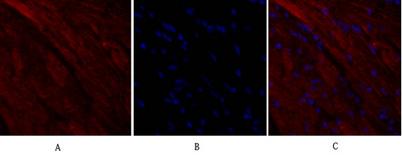
Immunofluorescence analysis of mouse-heart tissue.1,OPG Polyclonal Antibody(red) was diluted at 1:200(4°C,overnight). 2, Cy3 labled Secondary antibody was diluted at 1:300(room temperature, 50min).3, Picture B: DAPI(blue) 10min. Picture A:Target. Picture B: DAPI. Picture C: merge of A+B

No documents available.