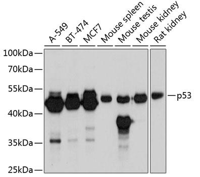
Western blot analysis of extracts of various cell lines, using p53 antibody  at 1:6000 dilution.
Secondary antibody: HRP Goat Anti-Rabbit IgG (H+L) at 1:10000 dilution.
Lysates/proteins: 25ug per lane.
Blocking buffer: 3% nonfat dry milk in TBST.
Detection: ECL West Pico Plus.
Exposure time: 15s.

Western blot analysis of extracts of various cell lines, using p53 antibody at 1:6000 dilution. Secondary antibody: HRP Goat Anti-Rabbit IgG (H+L) at 1:10000 dilution. Lysates/proteins: 25ug per lane. Blocking buffer: 3% nonfat dry milk in TBST. Detection: ECL West Pico Plus. Exposure time: 15s.

Catalog Number:AB-83528
Size:100 ug
Concentration:1mg/ml
Host:Rb
Isotype:IgG
Clone:POLY
Immunogen:A synthetic peptide corresponding to a sequence within amino acids 1-100 of human p53.
Reactivity:Hu, Ms, Rt
Applications:Western Blot: 1:3000 - 1:6000 Immunohistochemistry: 1:50 - 1:200 Immunoprecipitation: 1:20 - 1:50 ChIP: 1:20 - 1:50
Molecular Weight:53kDa
Purification:Aff. Pur.
Form:Liquid

Applications

Western Blot: 1:3000 - 1:6000 Immunohistochemistry: 1:50 - 1:200 Immunoprecipitation: 1:20 - 1:50 ChIP: 1:20 - 1:50

Immunogen

A synthetic peptide corresponding to a sequence within amino acids 1-100 of human p53.
Target Background

This gene encodes tumor protein p53, which responds to diverse cellular stresses to regulate target genes that induce cell cycle arrest, apoptosis, senescence, DNA repair, or changes in metabolism. p53 protein is expressed at low level in normal cells and at a high level in a variety of transformed cell lines, where it's believed to contribute to transformation and malignancy. p53 is a DNA-binding protein containing transcription activation, DNA-binding, and oligomerization domains. It is postulated to bind to a p53-binding site and activate expression of downstream genes that inhibit growth and/or invasion, and thus function as a tumor suppressor. Mice deficient for this gene are developmentally normal but are susceptible to spontaneous tumors. Evidence to date shows that this gene contains one promoter, in contrast to alternative promoters of the human gene, and transcribes a few of splice variants which encode different isoforms, although the biological validity or the full-length nature of some variants has not been determined.

Storage

Store at –20°C. Avoid Freeze and Thaw cycles.

Buffer

PBS with 0.02% sodium azide, 50% glycerol, pH7.3

Western blot analysis of extracts of various cell lines, using p53 antibody at 1:6000 dilution. Secondary antibody: HRP Goat Anti-Rabbit IgG (H+L) at 1:10000 dilution. Lysates/proteins: 25ug per lane. Blocking buffer: 3% nonfat dry milk in TBST. Detection: ECL West Pico Plus. Exposure time: 15s.

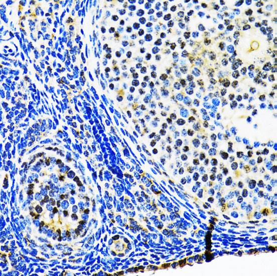
Immunohistochemistry of paraffin embedded embedded rat ovary using p53 antibody at dilution of 1:100 (40x lens).

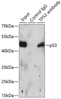
Immunoprecipitation analysis of 150ug extracts of A549 cells using 3ug p53 antibody. Western blot was performed from the immunoprecipitate using p53 antibod at a dilition of 1:500.

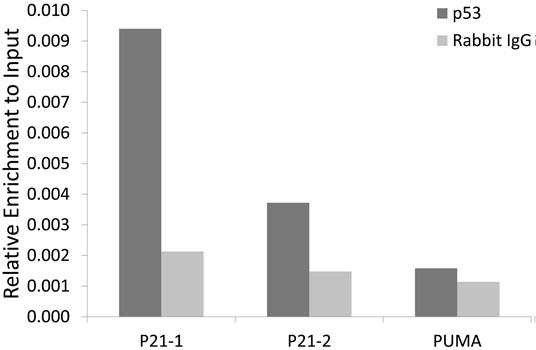
Chromatin immunoprecipitation analysis of extracts of 293T cells, using p53 antibody and rabbit IgG. The amount of immunoprecipitated DNA was checked by quantitative PCR. Histogram was constructed by the ratios of the immunoprecipitated DNA to the input.

No documents available.