p53 Di-Methyl- (Lys370) Rabbit Polyclonal Antibody

Western Blot analysis of MCF7, K562 cells using Di-Methyl-p53 (K370) Polyclonal Antibody. Secondary antibody  was diluted at 1:20000

Western Blot analysis of MCF7, K562 cells using Di-Methyl-p53 (K370) Polyclonal Antibody. Secondary antibody  was diluted at 1:20000

Catalog Number:AB-84047
Conjugate:Unconjugated
Size:100ug
Concentration:1mg/ml
Host:Rabbit
Isotype:IgG
Clone:POLY
Immunogen:Synthesized peptide derived from human p53 around the di-methylation site of K370.
Reactivity:Human;Rat;Mouse;
Applications:Western Blot: 1/500 - 1/2000 Immunohistochrmistry (paraffin-embedded tissues):1:100-300 Immunofluorescence:  1:50-200
Molecular Weight:53kD
Purification:The antibody was affinity-purified from rabbit antiserum by affinity-chromatography using epitope-specific immunogen.
Form:Liquid
Synonyms:TP53; P53; Cellular tumor antigen p53; Antigen NY-CO-13; Phosphoprotein p53; Tumor suppressor p53

Applications

Western Blot: 1/500 - 1/2000 Immunohistochrmistry (paraffin-embedded tissues):1:100-300 Immunofluorescence:  1:50-200

Immunogen

Synthesized peptide derived from human p53 around the di-methylation site of K370.
Target Background

tumor protein p53(TP53) Homo sapiens This gene encodes a tumor suppressor protein containing transcriptional activation, DNA binding, and oligomerization domains. The encoded protein responds to diverse cellular stresses to regulate expression of target genes, thereby inducing cell cycle arrest, apoptosis, senescence, DNA repair, or changes in metabolism. Mutations in this gene are associated with a variety of human cancers, including hereditary cancers such as Li-Fraumeni syndrome. Alternative splicing of this gene and the use of alternate promoters result in multiple transcript variants and isoforms. Additional isoforms have also been shown to result from the use of alternate translation initiation codons.

Synonyms:TP53; P53; Cellular tumor antigen p53; Antigen NY-CO-13; Phosphoprotein p53; Tumor suppressor p53

Storage

Store at -20°C. Avoid repeated freeze-thaw cycles.

Buffer

Liquid in PBS containing 50% glycerol, 0.5% BSA and 0.02% sodium azide.

Western Blot analysis of MCF7, K562 cells using Di-Methyl-p53 (K370) Polyclonal Antibody. Secondary antibody  was diluted at 1:20000

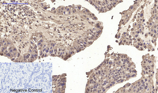
Immunohistochemical analysis of paraffin-embedded Human-liver-cancer tissue. 1,p53 (Di Methyl Lys370) Polyclonal Antibody was diluted at 1:200(4°C,overnight). 2, Sodium citrate pH 6.0 was used for antibody retrieval(>98°C,20min). 3,Secondary antibody was diluted at 1:200(room tempeRature, 30min). Negative control was used by secondary antibody only.

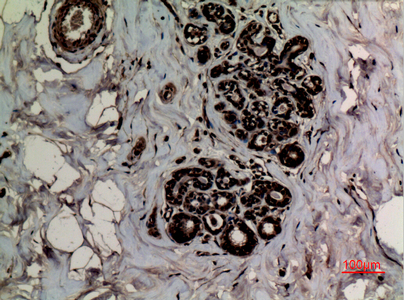
Immunohistochemical analysis of paraffin-embedded human-breast, antibody was diluted at 1:100

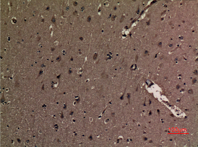
Immunohistochemical analysis of paraffin-embedded human-brain, antibody was diluted at 1:100

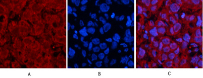
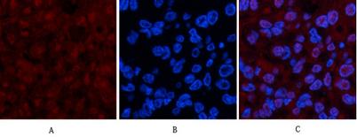
Immunofluorescence analysis of human-breast-cancer tissue. 1,p53 (Di Methyl Lys370) Polyclonal Antibody(red) was diluted at 1:200(4°C,overnight). 2, Cy3 labled Secondary antibody was diluted at 1:300(room temperature, 50min).3, Picture B: DAPI(blue) 10min

Immunofluorescence analysis of human-breast-cancer tissue. 1,p53 (Di Methyl Lys370) Polyclonal Antibody(red) was diluted at 1:200(4°C,overnight). 2, Cy3 labled Secondary antibody was diluted at 1:300(room temperature, 50min).3, Picture B: DAPI(blue) 10min. Picture A:Target. Picture B: DAPI. Picture C: merge of A+B

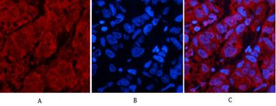
Immunofluorescence analysis of human-liver-cancer tissue. 1,p53 (Di Methyl Lys370) Polyclonal Antibody(red) was diluted at 1:200(4°C,overnight). 2, Cy3 labled Secondary antibody was diluted at 1:300(room temperature, 50min).3, Picture B: DAPI(blue) 10min.

No documents available.