Pan-Akt Rabbit mAb

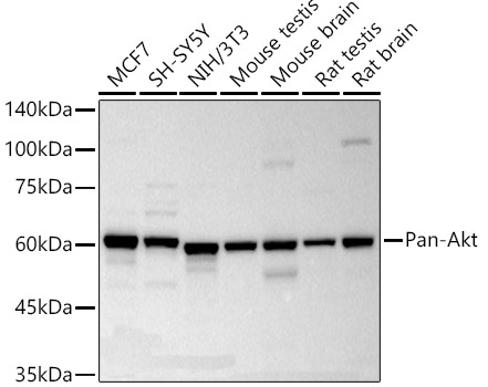
Western blot analysis of various lysates using Pan-Akt Rabbit mAb  at1:2000 dilution.Secondary antibody: HRP-conjugated Goat anti-Rabbit IgG   at1:10000 dilution.Lysates/proteins: 25μg per lane.Blocking buffer: 3% nonfat dry milk in TBST.Detection: ECL Basic Kit .Exposure time: 10s.

Western blot analysis of various lysates using Pan-Akt Rabbit mAb at1:2000 dilution.Secondary antibody: HRP-conjugated Goat anti-Rabbit IgG at1:10000 dilution.Lysates/proteins: 25μg per lane.Blocking buffer: 3% nonfat dry milk in TBST.Detection: ECL Basic Kit .Exposure time: 10s.

Catalog Number:AB18675
Conjugate:Unconjugated
Size:100 ug
Concentration:1mg/ml
Host:Rabbit
Isotype:IgG
Clone:ARC5005-05
Immunogen:Recombinant fusion protein containing a sequence corresponding to amino acids 1-123 of human Pan-Akt (NP_005154.2).
Reactivity:Human,Mouse,Rat
Applications:WB,1:2000 - 1:12000 IHC-P,1:200 - 1:800 IF/ICC,1:100 - 1:400 IP,0.5μg-4μg antibody for 200μg-400μg extracts of whole cells ELISA,Recommended starting concentration is 1 μg/mL. Please optimize the concentration based on your specific assay requirements.
Molecular Weight:60kDa
Purification:Affinity purification
Form:liquid
Synonyms:None

Human AKT serine-threonine protein kinase family includes three members AKT1,AKT2, AKT3, which are also often referred to as protein kinase B alpha, beta, and gamma. These highly similar AKT proteins all have an N-terminal pleckstrin homology domain, a serine/threonine-specific kinase domain and a C-terminal regulatory domain. These proteins are phosphorylated by phosphoinositide 3-kinase (PI3K). AKT/PI3K forms a key component of many signalling pathways that involve the binding of membrane-bound ligands such as receptor tyrosine kinases, G-protein coupled receptors, and integrin-linked kinase. These AKT proteins therefore regulate a wide variety of cellular functions including cell proliferation, survival, metabolism, and angiogenesis in both normal and malignant cells. AKT proteins are recruited to the cell membrane by phosphatidylinositol 3,4,5-trisphosphate (PIP3) after phosphorylation of phosphatidylinositol 4,5-bisphosphate (PIP2) by PI3K. Subsequent phosphorylation of both threonine residue 308 and serine residue 473 is required for full activation of the AKT1 protein encoded by this gene.

Applications

WB,1:2000 - 1:12000 IHC-P,1:200 - 1:800 IF/ICC,1:100 - 1:400 IP,0.5μg-4μg antibody for 200μg-400μg extracts of whole cells ELISA,Recommended starting concentration is 1 μg/mL. Please optimize the concentration based on your specific assay requirements.

Immunogen

Recombinant fusion protein containing a sequence corresponding to amino acids 1-123 of human Pan-Akt (NP_005154.2).

Target Background

Human AKT serine-threonine protein kinase family includes three members AKT1,AKT2, AKT3, which are also often referred to as protein kinase B alpha, beta, and gamma. These highly similar AKT proteins all have an N-terminal pleckstrin homology domain, a serine/threonine-specific kinase domain and a C-terminal regulatory domain. These proteins are phosphorylated by phosphoinositide 3-kinase (PI3K). AKT/PI3K forms a key component of many signalling pathways that involve the binding of membrane-bound ligands such as receptor tyrosine kinases, G-protein coupled receptors, and integrin-linked kinase. These AKT proteins therefore regulate a wide variety of cellular functions including cell proliferation, survival, metabolism, and angiogenesis in both normal and malignant cells. AKT proteins are recruited to the cell membrane by phosphatidylinositol 3,4,5-trisphosphate (PIP3) after phosphorylation of phosphatidylinositol 4,5-bisphosphate (PIP2) by PI3K. Subsequent phosphorylation of both threonine residue 308 and serine residue 473 is required for full activation of the AKT1 protein encoded by this gene.

Synonyms:None

Storage

Store at -20℃. Avoid freeze / thaw cycles.

Buffer

PBS with 0.05% proclin300,0.05% BSA,50% glycerol,pH7.3.

Western blot analysis of various lysates using Pan-Akt Rabbit mAb at1:2000 dilution.Secondary antibody: HRP-conjugated Goat anti-Rabbit IgG at1:10000 dilution.Lysates/proteins: 25μg per lane.Blocking buffer: 3% nonfat dry milk in TBST.Detection: ECL Basic Kit .Exposure time: 10s.

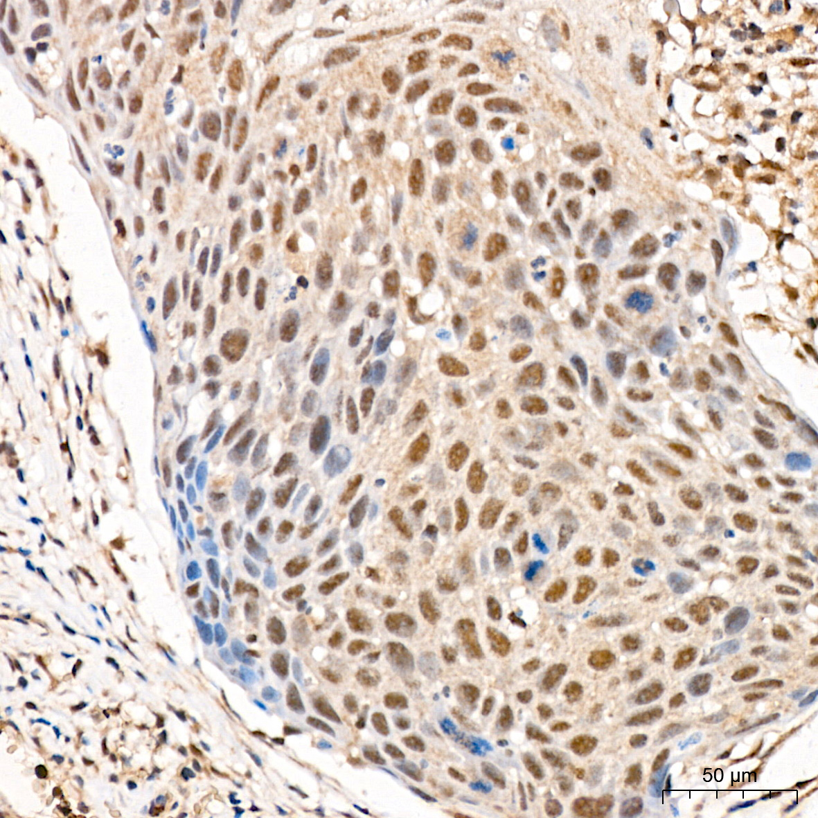
Immunohistochemistry analysis of paraffin-embedded Human cervix cancer tissue using Pan-Akt Rabbit mAb at a dilution of 1:200 . High pressure antigen retrieval performed with 0.01M Citrate Bufferr prior to IHC staining.

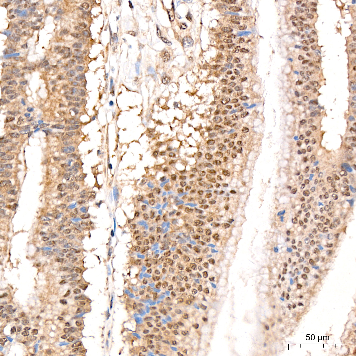
Immunohistochemistry analysis of paraffin-embedded Human colon carcinoma tissue using Pan-Akt Rabbit mAb at a dilution of 1:200 . High pressure antigen retrieval performed with 0.01M Citrate Bufferr prior to IHC staining.

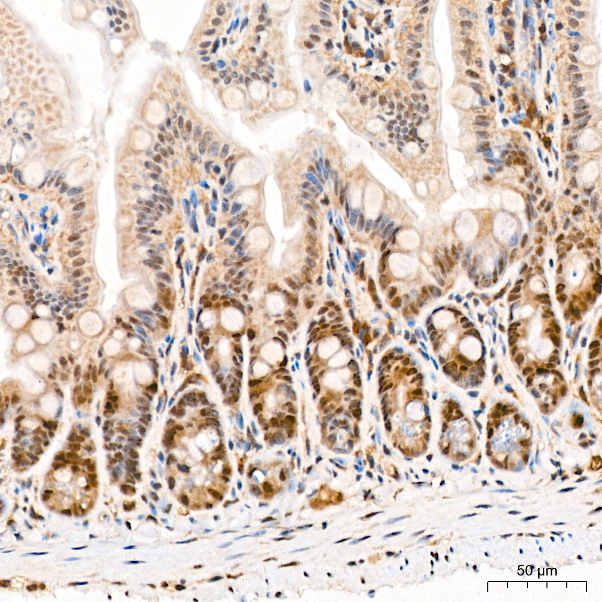
Immunohistochemistry analysis of paraffin-embedded Mouse intestin tissue using Pan-Akt Rabbit mAb at a dilution of 1:200 . High pressure antigen retrieval performed with 0.01M Citrate Bufferr prior to IHC staining.

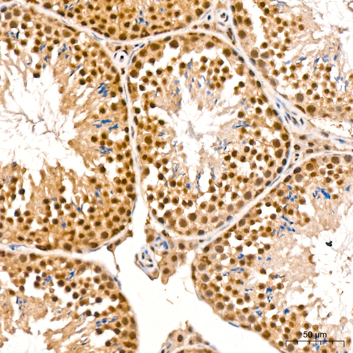
Immunohistochemistry analysis of paraffin-embedded Mouse testis tissue using Pan-Akt Rabbit mAb at a dilution of 1:200 . High pressure antigen retrieval performed with 0.01M Citrate Bufferr prior to IHC staining.

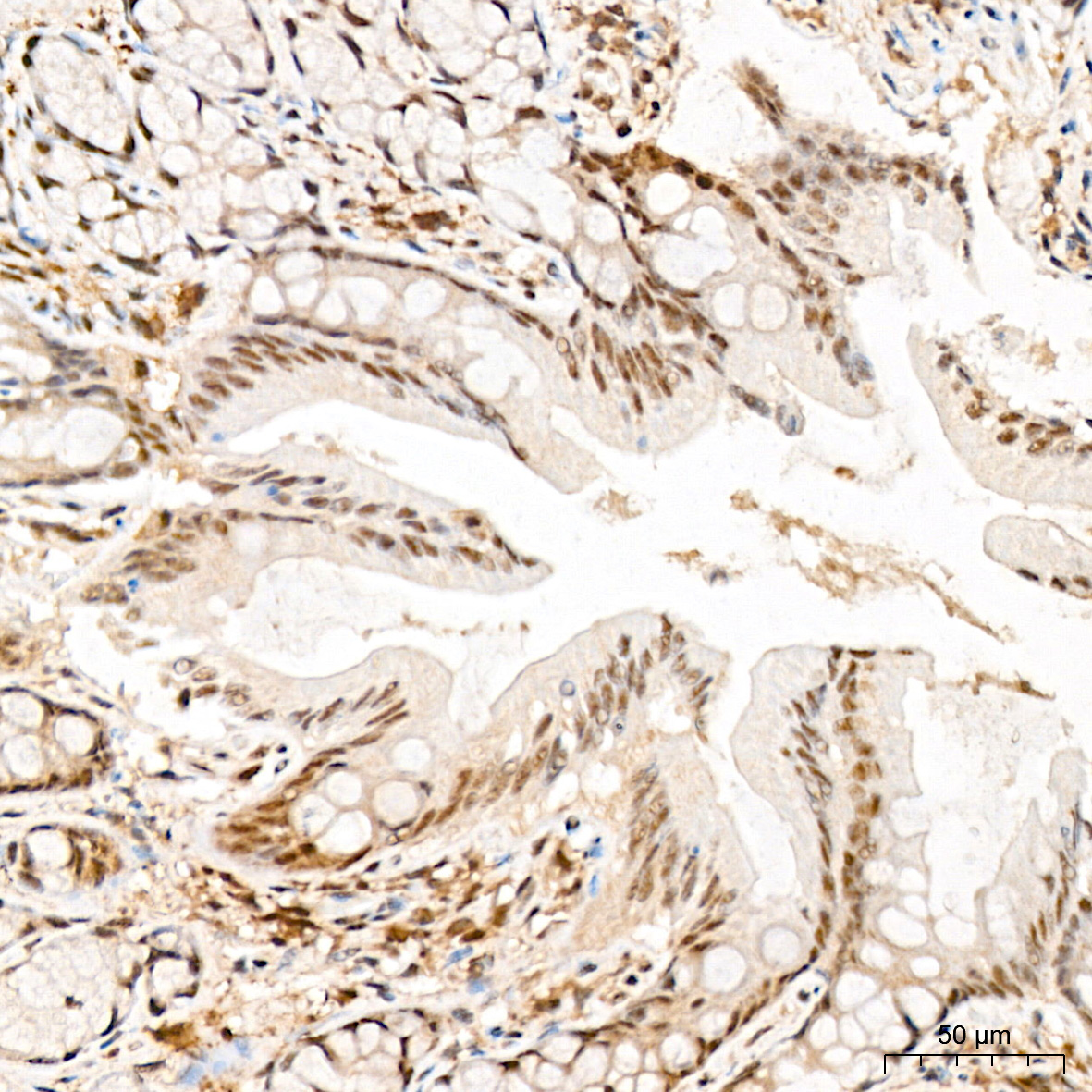
Immunohistochemistry analysis of paraffin-embedded Rat colon tissue using Pan-Akt Rabbit mAb at a dilution of 1:200 . High pressure antigen retrieval performed with 0.01M Citrate Bufferr prior to IHC staining.

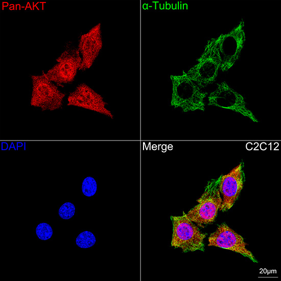
Confocal imaging of C2C12 cells using Pan-Akt Rabbit mAb followed by a further incubation with Cy3 Goat Anti-Rabbit IgG . The cells were counterstained with α-Tubulin Mouse mAb followed by incubation with ABflo® 488-conjugated Goat Anti-Mouse IgG Ab . DAPI was used for nuclear staining . Objective: 100x.

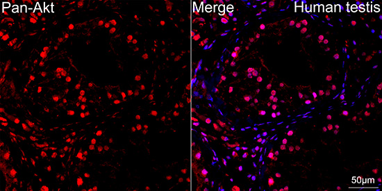
Confocal imaging of paraffin-embedded Human testis tissue using Pan-Akt Rabbit mAb followed by a further incubation with Cy3 Goat Anti-Rabbit IgG . DAPI was used for nuclear staining . High pressure antigen retrieval performed with 0.01M Citrate Buffer prior to IF staining. Objective: 40x.

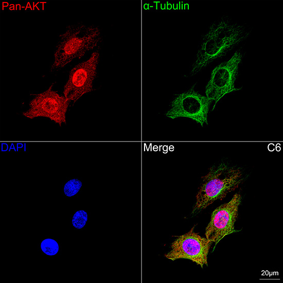
Confocal imaging of C6 cells using Pan-Akt Rabbit mAb followed by a further incubation with Cy3 Goat Anti-Rabbit IgG . The cells were counterstained with α-Tubulin Mouse mAb followed by incubation with ABflo® 488-conjugated Goat Anti-Mouse IgG Ab . DAPI was used for nuclear staining . Objective: 100x.

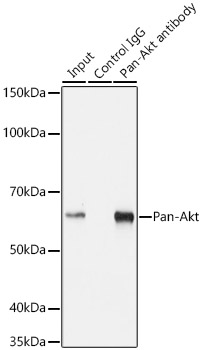
Immunoprecipitation analysis of 200 μg extracts from MCF7 cells using 3 μg Pan-Akt Rabbit mAb . Western blot was performed from the immunoprecipitate using Pan-Akt Rabbit mAb at a dilution of 1:1000.

No documents available.