
Pan Phospho-Threonine Rabbit mAb

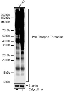
Western blot analysis of lysates from A-431, using Pan Phospho-Threonine Rabbit mAb (AP1422) at 1:1000 dilution. A 431 treated by Calyculin A (100 nM) at 37℃ for 30 minutes after serum-starvation overnight.
Secondary antibody: HRP-conjugated Goat anti-Rabbit IgG (H+L) (AS014) at 1:10000 dilution.
Lysates/proteins: 25μg per lane.
Blocking buffer: 3% nonfat dry milk in TBST.
Detection: ECL Enhanced Kit (RM00021).
Exposure time: 60s.

Western blot analysis of lysates from A-431, using Pan Phospho-Threonine Rabbit mAb (AP1422) at 1:1000 dilution. A 431 treated by Calyculin A (100 nM) at 37℃ for 30 minutes after serum-starvation overnight. Secondary antibody: HRP-conjugated Goat anti-Rabbit IgG (H+L) (AS014) at 1:10000 dilution. Lysates/proteins: 25μg per lane. Blocking buffer: 3% nonfat dry milk in TBST. Detection: ECL Enhanced Kit (RM00021). Exposure time: 60s.

Catalog Number:AB1422
Conjugate:Unconjugated
Size:100 ug
Concentration:1mg/ml
Host:Rabbit
Isotype:IgG
Clone:ARC61310
Immunogen:A synthetic phosphorylated peptide around of human Threonine.
Reactivity:Human,Mouse,Rat,Other (Wide Range Predicted)
Applications:WB,1:500 - 1:1000 ELISA,Recommended starting concentration is 1 μg/mL. Please optimize the concentration based on your specific assay requirements.
Molecular Weight:10-250kDa
Purification:Affinity purification
Form:liquid

Applications

WB,1:500 - 1:1000 ELISA,Recommended starting concentration is 1 μg/mL. Please optimize the concentration based on your specific assay requirements.

Immunogen

A synthetic phosphorylated peptide around of human Threonine.
Target Background

Storage

Store at -20℃. Avoid freeze / thaw cycles.

Buffer

PBS with 0.05% proclin300,0.05% BSA,50% glycerol,pH7.3.

Western blot analysis of lysates from A-431, using Pan Phospho-Threonine Rabbit mAb (AP1422) at 1:1000 dilution. A 431 treated by Calyculin A (100 nM) at 37℃ for 30 minutes after serum-starvation overnight. Secondary antibody: HRP-conjugated Goat anti-Rabbit IgG (H+L) (AS014) at 1:10000 dilution. Lysates/proteins: 25μg per lane. Blocking buffer: 3% nonfat dry milk in TBST. Detection: ECL Enhanced Kit (RM00021). Exposure time: 60s.

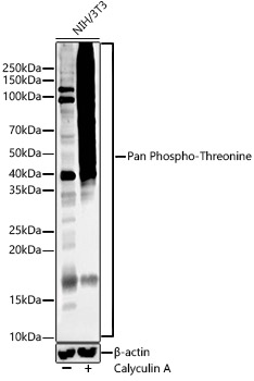
Western blot analysis of lysates from NIH 3T3 or NIH 3T3 treat with Calyculin A, using Pan Phospho-Threonine Rabbit mAb (AP1422) at 1:1000 dilution. NIH/3T3 cells were treated by Calyculin A (100 nM) at 37℃ for 30 minutes after serum-starvation overnight. Secondary antibody: HRP-conjugated Goat anti-Rabbit IgG (H+L) (AS014) at 1:10000 dilution. Lysates/proteins: 25μg per lane. Blocking buffer: 3% nonfat dry milk in TBST. Detection: ECL Enhanced Kit (RM00021). Exposure time: 60s.

No documents available.