PARK2

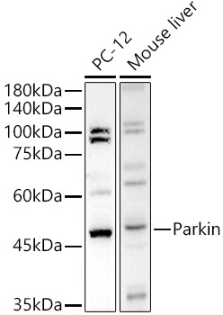
Western blot analysis of extracts of various cell lines, using Parkin antibody at 1:500 dilution.
Secondary antibody: HRP Goat Anti-Rabbit IgG (H+L) at 1:10000 dilution.
Lysates/proteins: 25μg per lane.
Blocking buffer: 3% nonfat dry milk in TBST.
Detection: ECL Basic Kit.
Exposure time: 180s

Western blot analysis of extracts of various cell lines, using Parkin antibody at 1:500 dilution. Secondary antibody: HRP Goat Anti-Rabbit IgG (H+L) at 1:10000 dilution. Lysates/proteins: 25μg per lane. Blocking buffer: 3% nonfat dry milk in TBST. Detection: ECL Basic Kit. Exposure time: 180s

Catalog Number:AB-84037
Conjugate:Unconjugated
Size:100ug
Concentration:1mg/ml
Host:Rabbit
Isotype:IgG
Clone:POLY
Immunogen:Recombinant fusion protein containing a sequence corresponding to amino acids 81-280 of human Parkin
Reactivity:Human, Mouse, Rat
Applications:Western Blot 1:100 - 1:500 Immunohistochemistry (P) 1:50 - 1:200
Molecular Weight:52 kDa
Purification:Affinity Purified
Form:Liquid
Synonyms:PDJ; AR-JP; LPRS2; PARKIN

Applications

Western Blot 1:100 - 1:500 Immunohistochemistry (P) 1:50 - 1:200

Immunogen

Recombinant fusion protein containing a sequence corresponding to amino acids 81-280 of human Parkin

Target Background

The precise function of this gene is unknown; however, the encoded protein is a component of a multiprotein E3 ubiquitin ligase complex that mediates the targeting of substrate proteins for proteasomal degradation. Mutations in this gene are known to cause Parkinson disease and autosomal recessive juvenile Parkinson disease. Alternative splicing of this gene produces multiple transcript variants encoding distinct isoforms. Additional splice variants of this gene have been described but currently lack transcript support.

Synonyms:PDJ; AR-JP; LPRS2; PARKIN

Storage

Store at -20℃. Avoid freeze / thaw cycles.

Buffer

PBS with 0.05% proclin300,50% glycerol, pH7.3.

Western blot analysis of extracts of various cell lines, using Parkin antibody at 1:500 dilution. Secondary antibody: HRP Goat Anti-Rabbit IgG (H+L) at 1:10000 dilution. Lysates/proteins: 25μg per lane. Blocking buffer: 3% nonfat dry milk in TBST. Detection: ECL Basic Kit. Exposure time: 180s

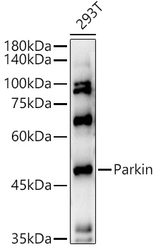
Western blot analysis of extracts of 293T cells, using Parkin antibody at 1:500 dilution. Secondary antibody: HRP Goat Anti-Rabbit IgG (H+L) at 1:10000 dilution. Lysates/proteins: 25μg per lane. Blocking buffer: 3% nonfat dry milk in TBST. Detection: ECL Enhanced Kit. Exposure time: 180s.

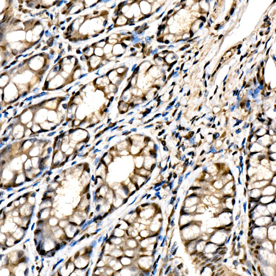
Immunohistochemistry analysis of paraffin-embedded mouse colon using Parkin Rabbit pAb at dilution of 1:100 (40x lens). Perform high pressure antigen retrieval with 10 mM citrate buffer pH 6.0 before commencing with IHC staining protocol

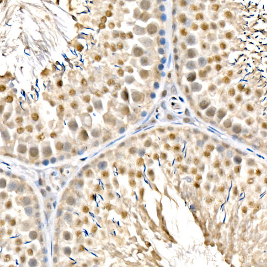
Immunohistochemistry analysis of paraffin-embedded rat testis using Parkin Rabbit pAb at dilution of 1:100 (40x lens). Perform high pressure antigen retrieval with 10 mM citrate buffer pH 6.0 before commencing with IHC staining protocol.

No documents available.